Houssam Halawi, MD, Thomas Stievenard, , Ayah Matar, MD, Jeffrey Fetzer, PhD, MS, Michael Camilleri, MD, DSc Mayo Clinic, Rochester, MN Introduction: Colonic volume is an important physiological and clinical parameter, especially in gastrointestinal motility disorders and functional bowel diseases. Various imaging modalities, such as computed tomography (CT), CT colonography, magnetic resonance imaging, barium enema and ultrasonography, can assess colonic volume and content. However, each technique has limitations. This study aimed to validate colonic volume measurements derived from scintigraphy performed during colonic transit assessments, compared with colonic volume obtained from contrast-enhanced CT scans. Methods: Following retrospective review of medical records, 39 patients who underwent both colonic scintigraphy and CT imaging in evaluation of chronic constipation were identified and were included for full colonic volume measurements using scintigraphy. Based on colonic visualization on scintigraphic transit study, colonic diameters and lengths on scintigraphy were measured manually by one investigator, tracing the central axis of the colonic lumen. Colonic volume on CT was quantified using “TotalSegmentator” software. Results were evaluated with Mann-Whitney test, Pearson correlation analysis and Bland-Altman analysis. Results: A total of 39 whole colons were analyzed (mean age: 52, 71.8% female sex). Median colonic transit at 48h was 2.8 (IQR 2.5-4.0), compared to normal median 4.52 (range 2.52-5.0) in 61 healthy adults. Colonic volumes were significantly lower with scintigraphy (median 660, IQR 483.7-829.4) than by CT (median 807, IQR 602.0-958.0 [p=0.044]), with an average difference of 126.4 mL (95% CI: –235.6 to –17.2). A Pearson correlation analysis showed a moderate positive correlation between CT and scintigraphic volumes (r = 0.528, p = 0.0006). Discussion: Although the difference in colonic volume between CT and scintigraphy is significant, the volumes measured by scintigraphy are significantly correlated with CT volumes. CT volume measurements were affected by segmentation variability and scan timing, while scintigraphic measurements were performed manually. Using a continuous central axis for length minimized overlap errors at segment transitions. Subjective diameter selection, non-blinding and the use of a cylindrical model could introduce bias. However, measuring colonic volume on scintigraphy during colonic transit demonstrated feasibility, and adds a physiologically relevant dimension that could be acquired during colonic transit measurement.
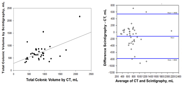
Figure: A) Moderate positive correlation between CT and scintigraphic colonic volumes (r = 0.528, p = 0.0006 using Pearson correlation analysis); B) Bland-Altman plot showing the degree of agreement between colonic volumes measured by scintigraphy and those measured using CT.
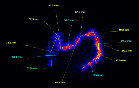
Figure: Example of colonic scintigraphy to assess colonic volume using a continuous central axis.