Sara C. Noguera, DO1, Tyler Doty, DO2, Jerome C. Edelson, MD3 1San Antonio Uniformed Services Health Education Consortium, Fort Sam Houston, TX; 2Brooke Army Medical Center, Cibolo, TX; 3Brooke Army Medical Center, San Antonio, TX Introduction: Schistosomiasis is a parasitic infection primarily affecting individuals in endemic regions such as Africa, Asia, and Latin America. While most infections are asymptomatic, chronic exposure can lead to significant pathology due to the host immune response to retained Schistosoma eggs. Eggs are transported via splanchnic circulation potentially embedding in various tissues, which can lead to granulomatous inflammation and fibrosis. In the colon, ulceration, bleeding, and polyp formation may occur; sometimes persisting years after successful treatment due to residual calcified ova and ongoing immune remodeling. This case highlights an unusual presentation of colonic polyps in an appropriately treated patient, underscoring the long-term sequelae of chronic intestinal schistosomiasis and the importance of considering it as a differential diagnosis, even in non-endemic settings.
Case Description/
Methods: A 67-year-old woman born in rural Philippines, now residing in the U.S., was diagnosed with hepatointestinal schistosomiasis in 2010 via liver biopsy during evaluation of abnormal liver function tests and chronic urticaria. She was treated with praziquantel (40 mg/kg, then 60 mg/kg six months later due to persistent symptoms). During a routine colonoscopy (CSP) in 2019 a sessile serrated adenoma and hyperplastic polyps were removed and showed evidence of calcified parasitic ova. Infectious disease (ID) deemed that no further treatment was required as she was asymptomatic and previously appropriately treated. On repeat screening CSP in 2024, a hyperplastic polyp (1) was resected and revealed calcified ova consistent with Schistosomiasis. Eosinophil counts remained consistently normal ( < 500/μL) without clinical suspicion for reinfection and the calcified ova was attributed to residual effects of a prior infection with S. japonicum. Once again, she was asymptomatic and no additional antiparasitic treatment was indicated at that time. Discussion: This case highlights an uncommon presentation of calcified Schistosoma ova identified in various polyp subtypes throughout the colon on CSP after appropriate treatment in an asymptomatic patient. While treatment with praziquantel eliminates Schistosoma worms, eggs are left untouched. Although these polyps are benign, they pose significant diagnostic challenges due to their resemblance to neoplastic lesions. There is limited but suggestive evidence for increased risk of colorectal carcinogenesis making awareness of clinical and pathological features essential.
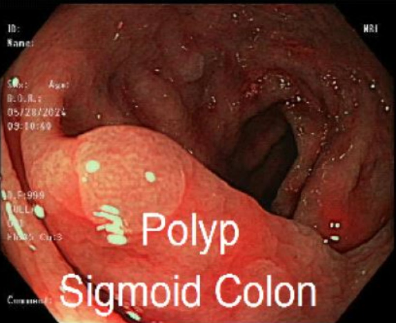
Figure: 3 mm sessile hyperplastic polyp found in the sigmoid colon
Disclosures: Sara Noguera indicated no relevant financial relationships. Tyler Doty indicated no relevant financial relationships. Jerome Edelson indicated no relevant financial relationships.
Sara C. Noguera, DO1, Tyler Doty, DO2, Jerome C. Edelson, MD3. P0372 - The Legacy Left Behind: Schistosomiasis Found in Sigmoid Colon Polyp in an Elderly Female, ACG 2025 Annual Scientific Meeting Abstracts. Phoenix, AZ: American College of Gastroenterology.