University of Texas Health, McGovern Medical School Houston, TX
Clay Smithhart, MD1, Tanvi Gupta, MD2, Aastha Bharwad, MD3, Ranya Selim, MD2 1University of Texas Health, McGovern Medical School, Houston, TX; 2University of Texas Health Science Center, Houston, TX; 3University of Texas at Houston, Houston, TX Introduction: Achalasia, an esophageal motility disorder marked by impaired peristalsis and relaxation of the lower esophageal sphincter, often presents with dysphagia and regurgitation. In rare cases, marked esophageal dilation may compress adjacent structures such as the left atrium or trachea, leading to hemodynamic compromise. Only isolated case reports describe such complications. We present a rare case of severe achalasia causing both tracheal and left atrial compression, posing significant procedural risks.
Case Description/
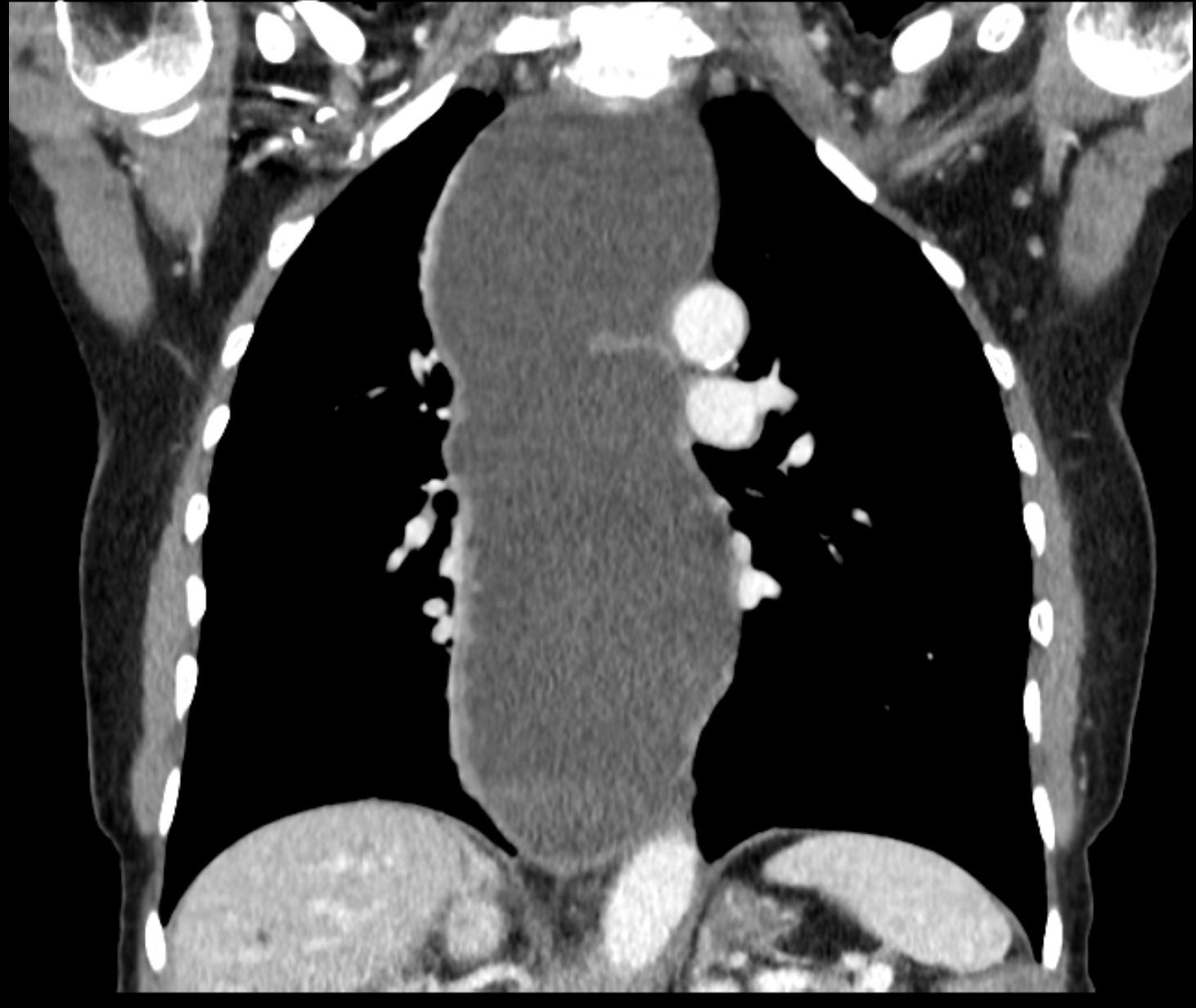
Methods: A 61-year-old uninsured woman presented to a county gastroenterology clinic with a 25-pound unintentional weight loss, dysphagia to solids and liquids, food regurgitation, and worsening chronic cough. Cross-sectional imaging showed severe distal esophageal distension with a possible mass at the gastroesophageal junction (GEJ) compressing the left atrium. Esophageal manometry revealed absent contractility and ineffective motility. Esophagogastroduodenoscopy (EGD) showed a tight GEJ stricture at 38cm with proximal esophageal distension, consistent with achalasia. Balloon dilation to 11.5mm and botulinum toxin injection (100 units) were performed. She was lost to follow up for over a year, and returned with persistent dysphagia, new onset exertional chest pain, and dyspnea. Transthoracic echocardiogram showed a large extracardiac mediastinal structure compressing the left atrium. Repeat cross-sectional imaging confirmed achalasia, now causing both tracheal and left atrial compression (figure 1). Cardiac stress test revealed abnormal hemodynamics and poor exercise tolerance. After multidisciplinary discussion with anesthesia, cardiology, and pulmonary, it was determined that she was at high risk for respiratory and circulatory collapse during sedation. Due to the significant procedural risks and the lack of advanced cardiopulmonary support capabilities at the county facility such as extracorporeal membrane oxygenation, the patient and family opted to pursue conservative management. Discussion: Massive esophageal dilation from achalasia may lead to life-threatening extrinsic compression of adjacent structures. This case demonstrates a rare finding of both tracheal and left atrial compression. Early recognition and timely intervention are crucial to avoid progression to a stage where interventions carry prohibitive risk. This case highlights the importance of multidisciplinary planning when evaluating these complex pathologies, particularly in resource-limited settings.
Figure: Cross-sectional imaging demonstrated severe esophageal distension, consistent with achalasia, compressing both the trachea and left atrium.
Disclosures: Clay Smithhart indicated no relevant financial relationships. Tanvi Gupta indicated no relevant financial relationships. Aastha Bharwad indicated no relevant financial relationships. Ranya Selim indicated no relevant financial relationships.
Clay Smithhart, MD1, Tanvi Gupta, MD2, Aastha Bharwad, MD3, Ranya Selim, MD2. P0702 - Severe Achalasia Causing Tracheal and Left Atrial Compression: A Multidisciplinary Challenge, ACG 2025 Annual Scientific Meeting Abstracts. Phoenix, AZ: American College of Gastroenterology.