Karmen Brar, MD1, Marianna Scranton, DO2 1University of Connecticut Health, Farmington, CT; 2Hartford HealthCare, Hartford, CT Introduction: Eosinophilic esophagitis (EoE) is a chronic, immune-mediated disease of the esophagus. It is seen with symptoms of esophageal dysfunction and histologic infiltration of ≥15 eosinophils per high-power field (eos/hpf) in the absence of other causes. Commonly recognized endoscopic features include rings, furrows, exudates and strictures. Here we present a unique case of EoE presenting with an endoscopic appearance of esophageal polypoid mucosa.
Case Description/
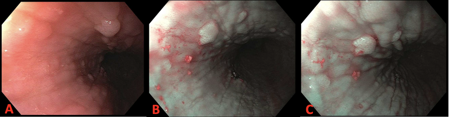
Methods: A 54-year-old woman presented with a one-year history of progressively worsening dysphagia, initially to solids eventually progressing to liquids. She denied associated heartburn, weight loss, or additional gastrointestinal symptoms. Upper endoscopy revealed severe esophagitis along with multiple polypoid lesions throughout the distal esophagus. Biopsies of polypoid mucosa revealed up to 101 eos/hpf within the esophageal epithelium, consistent with active EoE. Following initiation of high-dose PPI therapy, repeat endoscopy at two months showed significant mucosal healing, disappearance of polypoid mucosa, and near complete histologic resolution of eosinophilic infiltration. The patient’s dysphagia symptoms resolved in parallel with histologic improvement. Discussion: While classic endoscopic features include rings, furrows, exudates and strictures, polypoid mucosa is rarely reported and significantly broadens the known manifestations of EoE. Histopathology confirmed active EoE with >100 eos/hpf, absent dysplasia or viral cytopathic changes, supporting a reactive, eosinophil-driven inflammatory process. The polypoid appearance likely reflects localized eosinophilic micro-abscesses and chronic mucosal remodeling. Eosinophils release cytotoxic granules and profibrotic cytokines, leading to epithelial injury and a cycle of tissue regeneration that can result in nodular or polypoid growths. This is similar to pseudopolyps seen in inflammatory bowel disease. Similar findings have been noted in very few cases, with histologic confirmation of eosinophilic micro-abscesses within the polypoid mucosa. Most lesions in these cases resolved with standard EoE therapy, suggesting the changes are inflammatory rather than neoplastic. Our case reinforces that in rare instances, intense localized eosinophilic inflammation may produce polypoid mucosal projections as a form of mucosal remodeling. Awareness of this endoscopic variant is critical to avoid misdiagnosis and to recognize the full phenotypic spectrum of EoE.
Figure: Figure 1a. Upper Gastrointestinal tract. 1b-c., Middle third of esophagus.
Disclosures: Karmen Brar indicated no relevant financial relationships. Marianna Scranton indicated no relevant financial relationships.
Karmen Brar, MD1, Marianna Scranton, DO2. P0690 - Esophageal Polypoid Mucosa: An Atypical Presentation of EOE, ACG 2025 Annual Scientific Meeting Abstracts. Phoenix, AZ: American College of Gastroenterology.