Ibukunoluwa Oshobu, MD, MPH1, Gopala Konduri, MD2, Eunice Aregbesola, MD3, Nourhan Saleh, MD1, Hanna Nale, DO4, Ervin Tahan, 2, Muhammad Nadeem Yousaf, MD2 1University of Missouri Health Care, Columbia, MO; 2University of Missouri, Columbia, MO; 3University of Missouri, Columbia, Columbia, MO; 4University of Missouri School of Medicine, Columbia, MO Introduction: Hypopharyngeal lipomas are rare, benign tumors composed of mature adipose tissue located between the lower pharynx and proximal esophagus. While often asymptomatic, their pedunculated nature can lead to intermittent dysphagia, foreign body sensation, sleep apnea, airway obstruction, or stridor. In severe cases, they may mimic esophageal malignancy or cause sudden death if not recognized early.
Case Description/
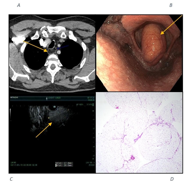
Methods: A 63-year-old man with a history of right-sided seminoma underwent a surveillance chest CT, which incidentally revealed an upper esophageal mass concerning for a giant esophageal polyp extending into the pharynx. The patient reported intermittent dysphagia to solids and semisolids, without odynophagia. He denied smoking or alcohol use. During EGD, the patient experienced an acute drop in oxygen saturation. On scope withdrawal to pharynx, a 20 mm pedunculated, polypoidal mass was visualized in the posterior hypopharynx, originating from the interarytenoid notch and impinging on the vocal cords. The airway was secured via endotracheal intubation and mass was re-evaluated endoscopically. EUS revealed a 26 x 17.8 mm isoechoic lesion with features suggestive of fatty infiltration. MRI of the face and neck showed a T1 hyperintense lesion consistent with lipoma. ENT performed a direct laryngoscopy with successful excisional biopsy. Histopathology demonstrated lobules of mature adipose tissue, positive for p16 and negative for HMB45 and A103, confirming lipoma. The patient had resolution of dysphagia and stable vocal function on follow-up. Discussion: Hypopharyngeal lipomas are a rare but important cause of intermittent or progressive dysphagia. This case highlights the “ball-valve” effect, where a mobile pedunculated mass intermittently obstructs the airway or esophageal inlet. Diagnosis requires high clinical suspicion, especially in patients with unexplained or positional symptoms. Gastroenterologists play a key role in identification and coordination with ENT for definitive diagnosis and surgical excision. Early recognition and multidisciplinary management are essential to prevent complications, including airway compromise. Resection is typically curative, with recurrence rates under 5%. This case also emphasizes the importance of securing the airway prior to endoscopic procedures in patients with known or suspected pharyngeal or esophageal masses to reduce the risk of aspiration or respiratory compromise during sedation.
Figure: FIGURE 1. A: CT scan showing fat density lesion in esophagus; B: EGD showing hypopharyngeal lipoma; C: EUS finding of hypopharyngeal lipoma; D: Hematoxylin and Eosin, 4x Lobules of Mature Adipose Tissue.
Disclosures: Ibukunoluwa Oshobu indicated no relevant financial relationships. Gopala Konduri indicated no relevant financial relationships. Eunice Aregbesola indicated no relevant financial relationships. Nourhan Saleh indicated no relevant financial relationships. Hanna Nale indicated no relevant financial relationships. Ervin Tahan indicated no relevant financial relationships. Muhammad Nadeem Yousaf indicated no relevant financial relationships.
Ibukunoluwa Oshobu, MD, MPH1, Gopala Konduri, MD2, Eunice Aregbesola, MD3, Nourhan Saleh, MD1, Hanna Nale, DO4, Ervin Tahan, 2, Muhammad Nadeem Yousaf, MD2. P0904 - A Ball Valve Effect of Hypopharyngeal Lipoma Causing Intermittent Dysphagia, ACG 2025 Annual Scientific Meeting Abstracts. Phoenix, AZ: American College of Gastroenterology.

 photo")