Lorraine Chong Tai, MD1, Edwin Makarevich, DO1, Ivana Rubenstein, DO1, Amanda Eukovich, DO1, Andrea Escalante, DO2, Ronen Arai, MD3 1Broward Health Medical Center, Fort Lauderdale, FL; 2Broward Health Medical Center, Miami, FL; 3Gastro Health, Coral Springs, FL Introduction: Patients with Inflammatory Bowel Disease (IBD) are at a 2 to 3-fold higher risk of developing a Venous Thromboembolism (VTE) as compared to the general population. We describe an interesting case of portal vein thrombosis as the first manifesting sign of Inflammatory Bowel Disease.
Case Description/
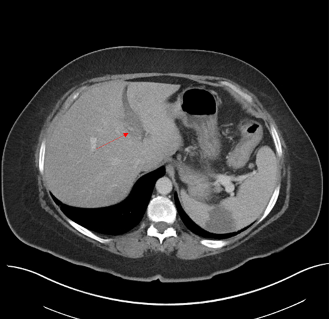
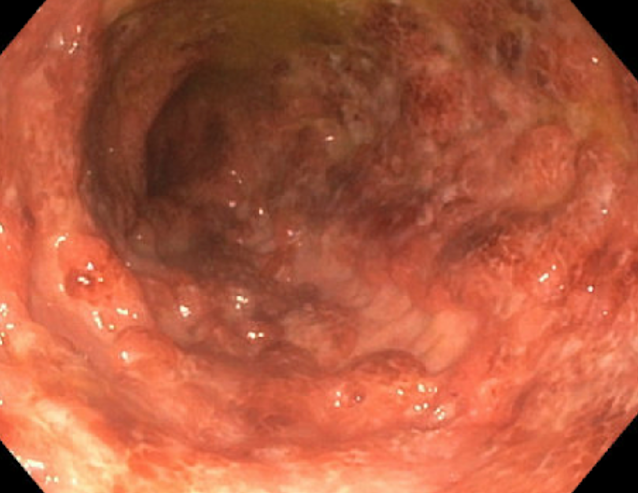
Methods: A 48-year-old female presented with a 2-week history of abdominal pain, nausea, vomiting and non-bloody diarrhea. She reported a similar episode 3 months prior that lasted for 3 weeks and resolved without medical attention at that time. A CT scan of the Abdomen and Pelvis showed Left portal vein thrombosis (Figure 1), diffuse colonic wall thickening with mild surrounding inflammatory changes, compatible with colitis and a wedge-shaped hypodensity within the spleen, compatible with splenic infarct. She denied any personal or family history of clotting disorders. She was started on anticoagulation with heparin and was initially started on antibiotics. Laboratory findings showed an elevated CRP and an infectious workup for her colitis was unremarkable. She later underwent a colonoscopy which showed severe diffuse colitis extending from the rectum to the proximal transverse colon (Figure 2). More proximal advancement was not performed due to severe ulceration, disease was much more severe in the proximal colon than the rectum. Biopsies showed lymphoplasmacytic infiltrate within the lamina propria, acute cryptitis, crypt abscess and focal ulceration. Given the severity of disease more proximal to the rectum, the clinical finding of non-bloody diarrhea and biopsy findings, Crohn’s disease was suspected. Antibiotics were subsequently discontinued. She was later discharged on prednisone and anticoagulation with Eliquis. At follow-up 1 week later, she endorsed some improvement in her symptoms with plans to start biologics for further treatment. Discussion: Portal vein thrombosis (PVT) has been shown to occur more frequently in patients with inflammatory bowel disease (IBD) compared to the general population. Studies have also found among these IBD patients, the complication was more often associated in the setting of recent abdominal surgery. Our case was particularly interesting given this patient was not diagnosed with IBD previously and this was her first presenting symptom. Nevertheless, this case underscores a potential association between PVT and IBD, highlighting the importance of maintaining a high index of suspicion for this complication within this patient population.
Figure: Figure 1: CT scan of abdomen and pelvis; red arrow points to the Left portal vein thrombus
Figure: Figure 1: Colonoscopy which showed severe diffuse colitis of the transverse colon