Scot M. Lewey, DO, FACG Banner Health, Fort Collins, CO Introduction: Gastrointestinal stromal tumors (GIST) are the most common of mesenchymal tumors in the gastrointestinal (GI) tract but can also arise outside of the GI tract in the abdomen or perirectal space. Such extragastrointestinal GISTs or E-GISTs are very rare, representing only 10% of GISTs and tend to occur in younger patients, be larger, more aggressive and be prone to recurrence. EGISTs of the perirectal space can be easily visualized and biopsied by rectal endosonogaphy (EUS).
Case Description/
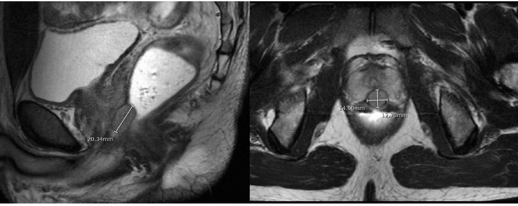
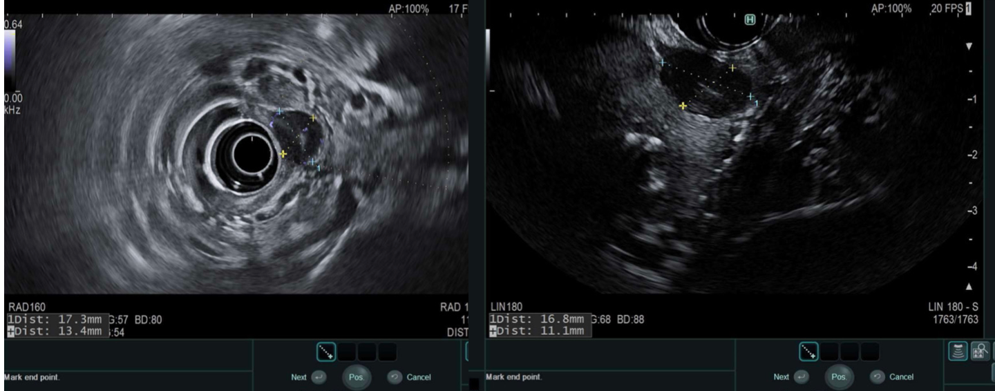
Methods: A 36-year-old man presented with symptoms of vague rectal fullness for two weeks without urinary or bleeding symptoms. A firm mass was noted on digital rectal exam. He was referred for colonoscopy that confirmed a submucosal lesion anteriorly in the rectum with normal overlying mucosa. Magnetic resonance imaging revealed a well demarcated 1.4 x 1.2 cm oval T1 isointense, T2 homogenously hypointense lesion with hypervascular enhancement at the anorectal junction between the prostate and rectum.Radial endosonography (EUS) revealed an oval 1.3 x 1.6 cm hypoechoic lesion with smooth margins in the anterior rectal space separate from the prostate gland. Curvilinear EUS was then performed allowing EUS guided fine needle core biopsy with a 22-gauge Franseen needle. Histologic and immunohistochemistry confirmed abundant groups of spindle cells that were positive for CD 117. The patient underwent preoperative imatinib therapy resulting in shrinkage of the lesion followed by uneventful trans-anal resection. Serial imaging revealed no recurrence over 24 months.
Discussion: EGISTs of the anterior rectal space are very rare. They may present with symptoms of rectal pain and fullness, urinary obstructive symptoms, vulvovaginal masses, or may be found during digital rectal or pelvic exams. Some are found pathologically after a prostate biopsy. They commonly occur in the anterior rectal space involving or separate from the prostate or vagina. Primary prostate GISTs have been reported but these arise within the prostate, not separate from it, as in this patient. Surgical excision is recommended with either preoperative or postoperative imatinib, but high local recurrence rates have been reported. This case illustrates the successful utilization of EUS to visualize and confirm a perirectal EGIST that then was successfully treated with imatinib chemotherapy followed by trans-anal resection.
Figure: MRI Images of Perirectal GIST
Figure: EUS Images of Perirectal GIST
Disclosures: Scot Lewey indicated no relevant financial relationships.
Scot M. Lewey, DO, FACG. P1459 - Endosonographic (EUS) FIne Needle Biopsy of an Extragastrointestinal Gastrointestinal Stromal Tumor (GIST) of the Perirectal Space, ACG 2025 Annual Scientific Meeting Abstracts. Phoenix, AZ: American College of Gastroenterology.