One Brooklyn Health-Brookdale University Hospital Medical Center Brooklyn, NY
Abdulrahman Atasi, MD1, Chidiebele Omaliko, MD1, Yasir Ali, MD1, Nader Daoud, MD2, Fatma Lina Emiroglu Atasi, MD3, Derrick Cheung, MD2 1One Brooklyn Health-Brookdale University Hospital Medical Center, Brooklyn, NY; 2Brookdale University Hospital Medical Center, Brooklyn, NY; 3Zucker School of Medicine-Long Island Jewish Medical Center, Hempstead, NY Introduction: Hepatocellular carcinoma (HCC) usually metastasizes through direct invasion, lymphatic route or by hematogenous dissemination. The most common sites are lungs, lymph nodes, and bones. Gastric metastasis from HCC is rare and usually occurs by direct invasion; hematogenous spread is rare. This case highlights HCC’s atypical gastric metastasis while imaging was suggesting metastatic liver disease with no evidence of direct invasion of the liver masses to the stomach.
Case Description/
Methods: A 70-year-old male with a history of alcohol use disorder, chronic kidney disease, and benign prostate hyperplasia who presented with dizziness, weakness and was admitted for symptomatic anemia. Initial laboratory work-up showed a hemoglobin of 5.9 g/dL which improved after blood transfusion. Abdominal ultrasound revealed markedly heterogeneous liver parenchyma most consistent with diffuse hepatic metastases. Further imaging with Magnetic Resonance of the abdomen with contrast identified innumerable, varying sized, solid masses compatible with diffuse extensive hepatic metastatic disease, irregular gastric wall thickening and ascites with findings suggestive of intraperitoneal carcinomatosis. Patient had no history of cirrhosis or imaging suggestive of cirrhosis. Due to imaging findings and presentation gastric cancer with liver metastasis was suspected, but serum alpha-fetoprotein was markedly elevated at 59,835 ng/mL, hepatitis B surface antigen and Hepatitis C virus antibody were nonreactive. Esophagogastroduodenoscopy demonstrated a friable, necrotic gastric mass extending from the proximal antrum through the gastric body, incisura and fundus, tissue biopsies from the mass were obtained. Histopathology of the gastric mass revealed invasive poorly differentiated carcinoma, with immunohistochemical stains consistent with high grade HCC involving the stomach. Likewise, a liver biopsy confirmed poorly differentiated HCC. Discussion: To this date, it is well-known for gastric cancers to metastasize to the liver, and present as multiple nodules in the liver parenchyma. However, not many case reports have been noted in the literature with a reversed phenomenon, a hepatocellular carcinoma metastasizing to the stomach. The case we are presenting is significant as imaging was suggestive of hepatic metastases with no history of cirrhosis or hepatitis. However, alpha-fetoprotein levels were significantly elevated which raised suspicion of HCC. Further testing revealed the diagnosis of HCC with a gastric metastasis.
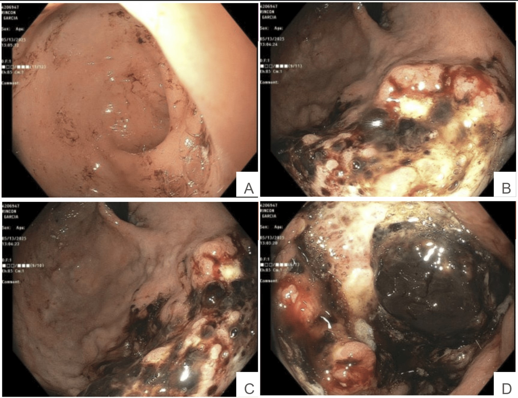
Figure: Images from Esophagogastroduodenoscopy showing a friable, necrotic gastric mass. A: Pre-pyloric stomach B: Gastric cardia C-D: Gastric body
Figure: Magnetic Resonance of the abdomen with contrast shows innumerable, varying sized, solid masses in the liver
Disclosures: Abdulrahman Atasi indicated no relevant financial relationships. Chidiebele Omaliko indicated no relevant financial relationships. Yasir Ali indicated no relevant financial relationships. Nader Daoud indicated no relevant financial relationships. Fatma Lina Emiroglu Atasi indicated no relevant financial relationships. Derrick Cheung indicated no relevant financial relationships.
Abdulrahman Atasi, MD1, Chidiebele Omaliko, MD1, Yasir Ali, MD1, Nader Daoud, MD2, Fatma Lina Emiroglu Atasi, MD3, Derrick Cheung, MD2. P1719 - When Hepatocellular Carcinoma Travels: Gastric Metastasis from Multifocal Hepatocellular Carcinoma - A Case Report, ACG 2025 Annual Scientific Meeting Abstracts. Phoenix, AZ: American College of Gastroenterology.

 photo")