Cleveland Clinic Abu Dhabi Abu Dhabi, Abu Dhabi, United Arab Emirates
Doa'a Alkhader, MD, Shahd Elhaj, MD, Kailash Makhejani, MD Cleveland Clinic Abu Dhabi, Abu Dhabi, Abu Dhabi, United Arab Emirates Introduction: Hepatic epithelioid hemangioendothelioma (HEHE) is a rare vascular liver malignancy, comprising < 1% of hepatic tumors, with a prevalence under 1 in 10 million. It affects all age groups, more commonly females, with a mean onset at 41.7 years. Diagnosis requires histopathology with immunohistochemical confirmation. Management varies based on disease burden, ranging from surveillance to liver transplantation. We present a case of multifocal HEHE in a young woman, highlighting individualized, multidisciplinary care.
Case Description/
Methods: A 29-year-old woman presented with intermittent abdominal discomfort. She denied systemic symptoms. Physical exam and labs, including liver enzymes, CBC, coagulation profile, and tumor markers (AFP, CEA, CA 19-9), were normal.
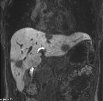
Ultrasound showed multiple hypoechoic liver lesions. Contrast-enhanced MRI revealed multifocal, peripheral-enhancing lesions with capsular retraction in both lobes (Figure 1). Differential included metastases, cholangiocarcinoma, or atypical infection.
Liver biopsy showed epithelioid endothelial cells in fibromyxoid stroma with intracytoplasmic vacuoles. Immunohistochemistry was positive for CD31, CD34, and factor VIII, confirming HEHE (Figure 2). PET-CT showed no extrahepatic spread or significant metabolic activity.
Due to multifocal unresectable disease, liver transplantation was recommended. After obtaining a second opinion, the patient chose surveillance. She was counseled on systemic options, including mTOR inhibitors (e.g., sirolimus), VEGF inhibitors (e.g., bevacizumab), and chemotherapy agents such as interferon-alpha, 5-FU, and doxorubicin, which show variable success in limited studies.
At 12-month follow-up, she remained asymptomatic with stable imaging and normal liver function. Discussion: HEHE often presents incidentally or with vague symptoms. Imaging findings are suggestive but not specific; biopsy is essential for diagnosis. Treatment strategies range from liver transplantation and systemic therapies—including mTOR inhibitors, interferon-alpha, VEGF inhibitors, and anthracycline-based chemotherapy—to surveillance in indolent cases. This case underscores the role of shared decision-making in managing this rare, unpredictable malignancy.
Figure: Figure (1): Coronal T2-weighted MRI of the liver demonstrating multiple hyperintense lesions consistent with hepatic epithelioid hemangioendothelioma (HEHE), predominantly involving both lobes with a peripheral distribution.
Figure: Figure (2) (A,B&C) Foci of low cellularity with myxohyaline and fibrotic stroma next to unremarkable liver tissue. These fibrotic foci showing small groups and single cells of epithelioid, stellate, spindle with fine chromatin with eosinophilic cytoplasm with occasional intracytoplasmic vacuoles ("blister cells"). No significant increase in mitosis. (D&E) The Immunohistochemical stains show these atypical epithelioid cells are expressing ERG and CAMTA1 (focal).
Disclosures: Doa'a Alkhader indicated no relevant financial relationships. Shahd Elhaj indicated no relevant financial relationships. Kailash Makhejani indicated no relevant financial relationships.
Doa'a Alkhader, MD, Shahd Elhaj, MD, Kailash Makhejani, MD. P1708 - Rare but Real: Hepatic Epithelioid Hemangioendothelioma Uncovered Through Vague Abdominal Pain, ACG 2025 Annual Scientific Meeting Abstracts. Phoenix, AZ: American College of Gastroenterology.

 photo")