University of Kansas School of Medicine Wichita, KS
Meagan H. Phox, DO1, Kevin J. Kadado, DO1, Mauricio Orrego, MD2 1University of Kansas School of Medicine, Wichita, KS; 2Advent Health Porter, Denver, CO Introduction: Portal vein stenosis (PVS) is a rare but recognized complication after liver transplantation (LT), potentially resulting in non-cirrhotic portal hypertension (HTN) and, in severe cases, graft failure. Doppler ultrasound (US) is the first-line imaging modality and highly sensitive for PVS. We report an unusual case of severe, late-onset PVS, diagnosed 13 years post-LT, which was missed on Doppler US and later identified via magnetic resonance imaging (MRI).
Case Description/
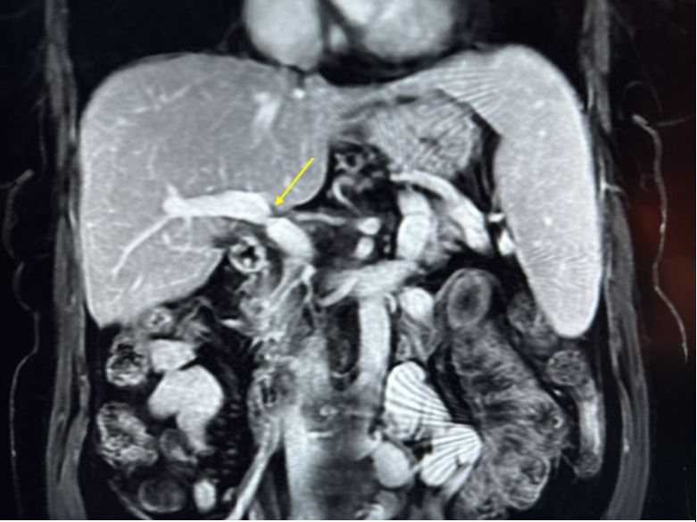
Methods: A 53-year-old female with a history of hepatitis C and alcoholic cirrhosis underwent LT in 2011 from a brain-dead donor. During her 2019 follow-up, US showed a nodular liver, inconsistent with non-invasive fibrosis testing that indicated F1-F2 fibrosis. Annual Doppler US suggested cirrhosis but remained stable. Esophagogastroduodenoscopy (EGD) revealed grade 1 esophageal varices (EV). Laboratory testing showed persistently mild alkaline phosphatase (ALP) elevation (~130 U/L), with aspartate aminotransferase and alanine aminotransferase mildly elevated at 28 U/L and 42 U/L, respectively. Mild thrombocytopenia was noted at 141,000/μL. Due to the nonspecific ALP elevation and imaging-lab discrepancies, MRI with and without contrast, and magnetic resonance cholangiopancreatography (MRCP) was performed. These revealed subtle cirrhotic features and significant PVS at the LT anastomosis(Figure 1). A diagnosis of non-cirrhotic portal HTN secondary to PVS was made. Interventional Radiology performed balloon angioplasty and a transjugular liver biopsy, which showed no steatosis, fibrosis, rejection, or active injury. A repeat EGD two months later showed complete resolution of EV. Discussion: This case illustrates a rare instance of PVS as a cause of prehepatic portal HTN emerging 13 years post-LT. The patient’s clinic status improved following angioplasty, demonstrated by EV resolution. This underscores the importance of recognizing delayed-onset vascular complications after LT. While Doppler US is a valuable first-line tool, its limitations may delay diagnosis. In patients with unexplained portal HTN and persistent liver abnormalities, cross-sectional imaging should be considered to avoid missed vascular pathology.
Figure: Figure 1. Coronal view of MRI Abdomen showing severe portal vein stenosis at the anastomosis site (arrow) from liver transplantation.
Disclosures: Meagan Phox indicated no relevant financial relationships. Kevin Kadado indicated no relevant financial relationships. Mauricio Orrego indicated no relevant financial relationships.
Meagan H. Phox, DO1, Kevin J. Kadado, DO1, Mauricio Orrego, MD2. P1792 - Atypical Presentation of Portal Vein Stenosis: Diagnosis 13 Years Post-Liver Transplantation, ACG 2025 Annual Scientific Meeting Abstracts. Phoenix, AZ: American College of Gastroenterology.

 photo")