Hospital Roosevelt / Gastri-k Guatemala City, San Marcos, Guatemala
Award: ACG Presidential Poster Award
Victoria Morales, 1, Kevin Molina, 2, Yuli Toledo, 3, Abel Sanchez, MD, MSc4 1Gastroenterology and Digestive Endoscopy, Roosevelt Hospital, Guatemala., Guatemala City, Chimaltenango, Guatemala; 2Gastroenterology and Digestive Endoscopy Unit, Hospital Roosevelt, Guatemala, Guatemala City, Chimaltenango, Guatemala; 3Gastroenterology an Digestive Endoscopy, Hospital Roosevelt, Guatemala, Guatemala City, Chimaltenango, Guatemala; 4Hospital Roosevelt / Gastri-k, Guatemala City, San Marcos, Guatemala Introduction: Gastrointestinal stromal tumors (GISTs) represent less than 1% of all gastrointestinal tumors. The most common location is the stomach (56%), followed by the small intestine (32%). Symptoms include dyspepsia, early satiety or upper gastrointestinal bleeding (UGIB). Small intestinal tumors with hemodynamic instability require urgent diagnosis and treatment. We present an 86-year-old male patient who presented with UGIB, showing a subepithelial tumor in the proximal jejunum with resected surgical specimen and adequate evolution
Case Description/
Methods: An 86-year-old Guatemalan man presented with a four-day history of melena. Hypotension, tachycardia (125 bpm), and generalized pallor were evident. Laboratory data revealed Hb in 3.1 mg/dl and elevated urea nitrogen 65 mg/dL. The patient was stabilized. Esophagogastroduodenosocopy was performed and no abnormalities. A colonoscopy, blood traces were observed in the colon and terminal ileum suggesting small bowel bleeding. Antegrade double-balloon enteroscopy was performed revealing a submucosal tumor measuring approximately 35 mm with a central depression, located in proximal jejunum. The patient persisted with active UGIB, performing his respective resuscitation and a consultation was requested to the Surgery Department. An exploratory laparotomy was performed with resection of a tumor in the proximal jejunum of 70 grams with measurements of 12.5 x 2.0 x 2.0 cm. The histopathological study confirmed stromal tumor, spindle cell type G1, low grade with KIT CD117+ and C34+ respectively. During follow-up, the patient was evaluated by oncology, who indicated conservative treatment. Patient discharged stable and without complications Discussion: GISTs located in the jejunum are rare, representing 0.1–3% of all gastrointestinal tumors. A few cases present with melena or hematemesis and severe anemia due to recurrent bleeding, causing hemodynamic instability and can lead to a life-threatening emergency if not diagnosed and treated promptly. In this case, due to the persistence of active bleeding and the urgent need to characterize the lesion, abdominal CT angiography and double-balloon enteroscopy were performed for timely characterization
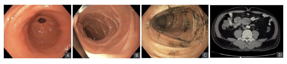
Figure: (A, B) no abnormalities or blood remnants in stomach and duodenum. (C) colonoscopy with blood remnants. D CT angiography (D) showed thickening of the proximal jejunum
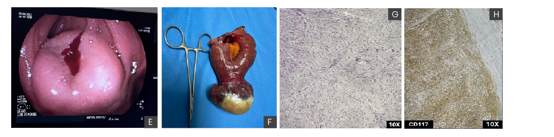
Figure: (E) Antegrade double-balloon enteroscopy with a submucosal tumor located in proximal jejunum (F) Surgical specimen of gastrointestinal stromal tumor. Pathological findings (G) Hematoxylin-Eosin (H&E) 10x irregularly enlarged spindle cells in bundles (H) Diffuse and moderate CD117 cytoplasmic immunoreactivity, 100×.
Disclosures: Victoria Morales indicated no relevant financial relationships. Kevin Molina indicated no relevant financial relationships. Yuli Toledo indicated no relevant financial relationships. Abel Sanchez indicated no relevant financial relationships.
Victoria Morales, 1, Kevin Molina, 2, Yuli Toledo, 3, Abel Sanchez, MD, MSc4. P1945 - Hemorrhagic Shock Secondary to a GIST Tumor in the Proximal Jejunum Diagnosed by Double-Balloon Enteroscopy, Angiography, and Laparoscopy, ACG 2025 Annual Scientific Meeting Abstracts. Phoenix, AZ: American College of Gastroenterology.


 photo")