Trevor McCracken, MD1, Andy Lin, MD2, Bryant Le, MD2, Marie Balfour, MD1, Kuangda Shan, MD3, Nathan Park, MD3, Peter Nguyen, MD2, Jason Samarasena, MD, MBA, FACG2, Elliot Yu, MD4, Robert Fearn, MD1 1University of California Irvine Health, Orange, CA; 2University of California Irvine, Orange, CA; 3University of California Irvine Digestive Health Institute, Orange, CA; 4University of California Irvine, Irvine, CA Introduction: Autoimmune enteropathy is a rare disorder characterized by chronic diarrhea, malabsorption, and villous atrophy, often presenting in children, although it may be seen in adults. Symptoms can mimic other causes of villous atrophy (e.g., Celiac disease), making timely diagnosis and management particularly challenging.
Case Description/
Methods: A 44-year-old female with vitiligo presented to a community emergency department with chronic diarrhea for three months associated with 40 pounds weight loss and severe hypokalemia. Initial studies revealed fecal elastase of 69 µg/g suggestive of exocrine pancreatic insufficiency, however, pancreatic enzyme replacement failed to improve her symptoms. Upper and lower GI endoscopy revealed a scalloped duodenum and was otherwise unremarkable.Biopsies demonstrated gastritis and enteritis with severe villous atrophy throughout the duodenum, jejunum, and terminal ileum. All colonic biopsies were unremarkable. After transfer to an academic medical center extensive testing was performed including celiac serologies, HLA DQ2 and DQ8, 24-hour urine 5-HIAA, serum VIP, serum gastrin, and serum chromogranin, GI PCR, C. diff, stool culture, stool ova and parasites, HIV, ASCA – all of which were negative. Calprotectin was minimally elevated at 51µg/g. 24-hour fecal alpha-1 antitrypsin clearance was elevated suggestive of a protein losing enteropathy. Repeat endoscopic evaluation redemonstrated severe villous atrophy, diminished goblet cells and Paneth cells, intraepithelial lymphocytosis, increased crypt epithelial apoptosis and acute on chronic inflammation throughout the duodenum, jejunum and terminal ileum. Random colon biopsy with markedly increased crypt epithelial apoptosis and acute inflammation. Expert pathologist review indicated that the findings were most suggestive of autoimmune enteropathy pattern of injury. The patient was started on empiric budesonide with prompt symptom improvement and is pending a repeat endoscopic evaluation after steroid taper for histologic resolution. Discussion: Autoimmune enteropathy in adults is a rare condition, and due to the lack of highly sensitive confirmatory tests, diagnosis can be challenging. A strong clinical suspicion is essential for identifying this disorder.
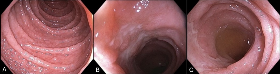
Figure: Figure 1. Diffuse nodularity with areas of villous blunting in the duodenum (A), terminal ileum with villous blunting and area of mucus that could not be washed off (B), terminal ileum with villous atrophy and ulceration (C).
Disclosures: Trevor McCracken indicated no relevant financial relationships. Andy Lin indicated no relevant financial relationships. Bryant Le indicated no relevant financial relationships. Marie Balfour indicated no relevant financial relationships. Kuangda Shan indicated no relevant financial relationships. Nathan Park indicated no relevant financial relationships. Peter Nguyen indicated no relevant financial relationships. Jason Samarasena: Applied Medical – Consultant. Boston Scientific – Consultant. Cook Medical – Consultant. Neptune Medical – Consultant. Olympus – Consultant. Elliot Yu indicated no relevant financial relationships. Robert Fearn indicated no relevant financial relationships.
Trevor McCracken, MD1, Andy Lin, MD2, Bryant Le, MD2, Marie Balfour, MD1, Kuangda Shan, MD3, Nathan Park, MD3, Peter Nguyen, MD2, Jason Samarasena, MD, MBA, FACG2, Elliot Yu, MD4, Robert Fearn, MD1. P2012 - The Disappearing Villi: Unmasking Autoimmune Enteropathy in a Mysterious Case of Chronic Diarrhea, ACG 2025 Annual Scientific Meeting Abstracts. Phoenix, AZ: American College of Gastroenterology.