McLaren Greater Lansing Hospital Dearborn Heights, MI
Ali Rida, MD1, Alexander Peskin, BS2, Claire C. Russell, DO3, Caleb Glover, DO3, Thomas Barker, MD4, Edward Cay, DO5, Eric Nguyen, MD3, Jannel Lee-Allen, MD3 1McLaren Greater Lansing Hospital, Dearborn Heights, MI; 2Michigan State University College of Osteopathic Medicine, Lansing, MI; 3McLaren Greater Lansing Hospital, Lansing, MI; 4Michigan State University, East Lansing, MI; 5McLaren Greater Lansing, Lansing, MI Introduction: Metastatic disease to the small intestine is uncommon and often mimics primary gastrointestinal tumors, particularly when the overlying mucosa is involved. The duodenum is a rare site of metastasis, and lesions here may present with gastric outlet obstruction. This case describes an obstructing duodenal mass initially presumed to be a primary adenocarcinoma but later confirmed as metastatic disease from an unidentified primary tumor.
Case Description/
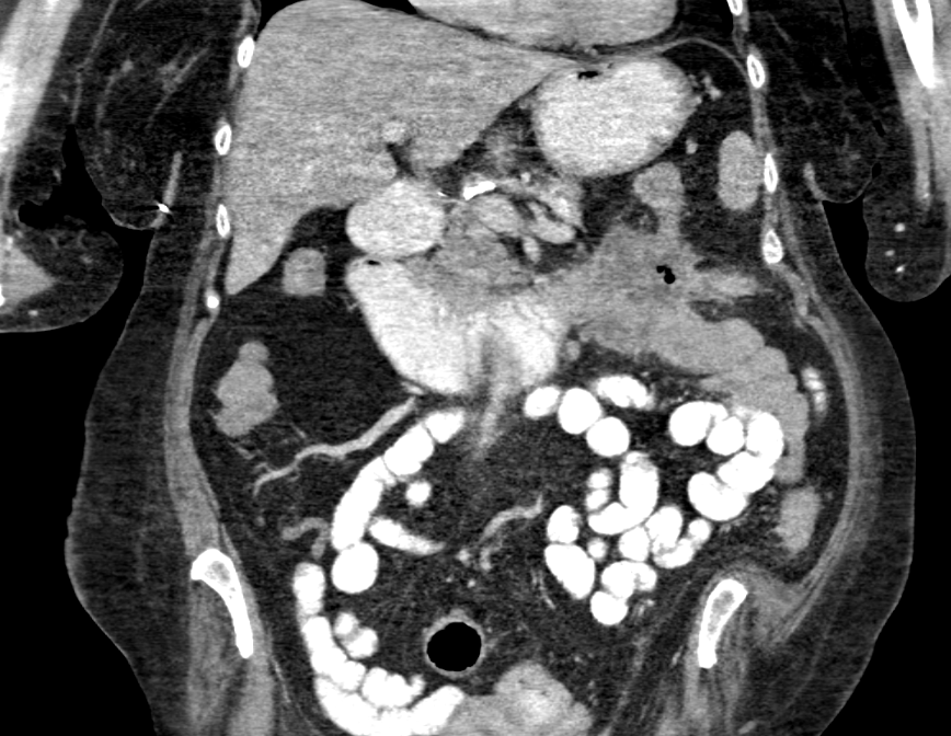
Methods: A 69-year-old woman with hypertension and chronic kidney disease presented with progressive weakness, postprandial vomiting, and two days of poor oral intake. Labs revealed hypokalemia, metabolic alkalosis, and mild azotemia. CT imaging showed a 6.1 × 5.0 × 4.7 cm mass in the third portion of the duodenum, encasing the superior mesenteric artery and abutting the ligament of Treitz, with adjacent mesenteric lymphadenopathy. Esophagogastroduodenoscopy demonstrated an ulcerated, partially obstructing mass. Biopsies revealed moderately differentiated adenocarcinoma beneath intact duodenal mucosa without dysplasia. Immunohistochemistry showed CK7-negative, CK20 patchy-positive, CDX2 positive, SATB2 weakly positive, and TTF-1 negative—suggesting a lower GI origin. Mismatch repair proteins (MLH1, MSH2, MSH6, PMS2) exhibited intact nuclear expression, indicating microsatellite stability. Extensive imaging, including PET-CT, failed to identify a primary tumor. The patient remained hospitalized for 37 days due to nutritional compromise and electrolyte derangements. Surgical resection was not feasible due to comorbidities and vascular encasement. Palliative chemotherapy was planned, but she was readmitted shortly after discharge with persistent vomiting. With declining functional status, she opted for hospice care. Discussion: Metastatic adenocarcinoma to the GI tract may closely resemble primary malignancy, particularly when mucosal colonization is present. In this case, the absence of epithelial dysplasia raised suspicion for a metastatic process. IHC findings were consistent with colorectal origin, while negative TTF-1 excluded pulmonary or gastric sources. Intact mismatch repair status ruled out Lynch syndrome. This case underscores the importance of integrating histology, IHC, and imaging to distinguish primary from metastatic GI lesions. Diagnostic accuracy is essential, as treatment strategies differ substantially—surgical resection for resectable primary disease versus systemic therapy or palliative interventions for metastases.
Figure: CT Imaging demonstrating multilobulated mass involving the the distal duodenum and proximal jejunum.
Figure: Small Bowel Enteroscopy imaging demonstrating fungating, protruding mass in duodenum
Disclosures: Ali Rida indicated no relevant financial relationships. Alexander Peskin indicated no relevant financial relationships. Claire Russell indicated no relevant financial relationships. Caleb Glover indicated no relevant financial relationships. Thomas Barker indicated no relevant financial relationships. Edward Cay indicated no relevant financial relationships. Eric Nguyen indicated no relevant financial relationships. Jannel Lee-Allen indicated no relevant financial relationships.
Ali Rida, MD1, Alexander Peskin, BS2, Claire C. Russell, DO3, Caleb Glover, DO3, Thomas Barker, MD4, Edward Cay, DO5, Eric Nguyen, MD3, Jannel Lee-Allen, MD3. P2009 - Metastatic Adenocarcinoma Masquerading as Primary Duodenal Cancer: A Case of Obstructive GI Lesion Without a Primary Source, ACG 2025 Annual Scientific Meeting Abstracts. Phoenix, AZ: American College of Gastroenterology.

 photo")