
Monday Poster Session
Category: Colon

Lefika Bathobakae, MD, MPH
St. Joseph's University Medical Center
Paterson, NJ
Endometrial adenocarcinoma is the most common gynecologic malignancy, with metastatic spread typically involving the lungs, liver, and distant lymph nodes. GI involvement, particularly rectal metastasis, is exceedingly rare and may mimic primary colorectal cancer or endometriosis. This overlap can lead to diagnostic uncertainty and delays in appropriate management. Herein, we present a novel case of endometrial adenocarcinoma with metastasis to the rectum and recto-uterine fistula, posing a significant diagnostic and therapeutic dilemma.
Case Description/
Methods:
A 58-year-old woman with a history of IDA presented to the ED after a syncopal episode at home. Although the patient denied any cardiac complaints in the ED, she reported a month history of intermittent vaginal bleeding. Triage blood tests were significant for low hemoglobin, 6.0 g/dL and reactive thrombocytosis, 525 K/mm3. EKG was negative for acute ischemia and troponin level was normal. The patient declined both digital rectal and pelvic examinations. CT of the abdomen and pelvis revealed rectosigmoid colitis with a suspected recto-uterine fistula. MRI of the pelvis showed a large rectal mass invading the uterus, with characteristics suggestive of a T4b N0 rectal neoplasm.
A colonoscopy demonstrated a fungating partially obstructing large mass in the rectum measuring 10 cm in length (Figure 1). A malignant-appearing, intrinsic stenosis was found in the rectum and traversed. Histopathology of the rectal mass showed colorectal mucosa with metastatic poorly differentiated adenocarcinoma, consistent with endometrial primary. Immunohistochemical analysis further supported the diagnosis, showing positivity for CK7, PAX8, and Vimentin. Stains for CDX2 and CK20 were negative. Despite a series of multidisciplinary conferences, the patient declined all interventions and was deemed to have medical capacity.
Discussion:
Metastatic endometrial adenocarcinoma to the rectum can be a challenging diagnosis due to its rarity and tendency to mimic primary GI cancers. Typically, metastatic endometrial adenocarcinoma cells are positive for CK7, estrogen receptor, and progesterone receptor and negative for CK20 and villin, which helps differentiate them from primary colorectal cancer.1 In this case, although the diagnostic imaging results were suggestive of primary rectal cancer, histopathology confirmed endometrial cancer, which has a unique therapeutic approach. Management usually involves surgical resection followed by adjuvant chemotherapy.2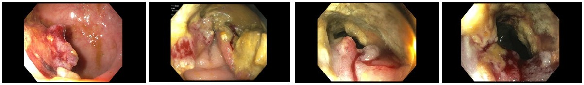
Figure: Figure 1. Endoscopy images showing a fungating, partially obstructing large mass in the rectum, measuring 10 cm in length, stricture in the rectum, and non-bleeding internal hemorrhoids. The bowel preparation was fair.
Disclosures:
Lefika Bathobakae indicated no relevant financial relationships.
Tasnim Fatima indicated no relevant financial relationships.
Abel Setlhoka indicated no relevant financial relationships.
Kajol Patel indicated no relevant financial relationships.
Nader Mekheal indicated no relevant financial relationships.
Mina Fransawy Alkomos indicated no relevant financial relationships.
Walid Baddoura indicated no relevant financial relationships.
Abraham El-Sedfy indicated no relevant financial relationships.
Lefika Bathobakae, MD, MPH1, Tasnim Fatima, MD1, Abel Setlhoka, BS2, Kajol Patel, MD3, Nader Mekheal, MD1, Mina Fransawy Alkomos, MD1, Walid Baddoura, MD1, Abraham El-Sedfy, MD, MSc1. P2484 - Endometrial Adenocarcinoma With Metastasis to the Rectum: A Diagnostic and Therapeutic Dilemma, ACG 2025 Annual Scientific Meeting Abstracts. Phoenix, AZ: American College of Gastroenterology.