Hasan Saleh, MD, MBA1, Salvatore Crucillà, 2, Jessica Petrov, MD, MSc1, Bhaumik Brahmbhatt, MBBS, MD1, Michele Lewis, MD1 1Mayo Clinic, Jacksonville, FL; 2University of Verona, Verona, Veneto, Italy Introduction: Ectopic pancreas (EP), or pancreatic rest, refers to pancreatic tissue located outside the usual anatomical location. EP is a congenital anomaly identified in 1–2% of autopsies and is usually asymptomatic.
Case Description/
Methods: A 47-year-old man with gastroesophageal reflux, peptic ulcer disease, and hepatic cysts presented with chronic episodic nausea, vomiting, abdominal pain, and weight loss. Laboratory tests showed a normal lipase level (34 U/L) and normal liver enzymes. MRI showed a 12.7 x 7.5 x 7.7 cm complicated fluid collection with a thick wall and internal debris in the right upper quadrant. Wall thickening of the gastric antrum and the first portion of the duodenum was present. Endoscopic ultrasound showed a 13mm by 16 mm submucosal nodule in the gastric antrum that was hypoechoic and contained anechoic tubular structures. A large anechoic cyst extended from the perigastric space close to the ectopic pancreas to the right upper quadrant gallbladder fossa. Fine needle aspiration of the cyst revealed amorphous debris, cloudy yellow fluid with high amylase (4897 U/L), low CEA (93 ng/mL), high lipase (16,700 U/L), and negative cytology, consistent with a pancreatic pseudocyst arising from EP tissue. The patient felt better after cyst aspiration and ultimately underwent an elective exploratory laparotomy involving excision of the cyst, distal gastrectomy of the EP, and Roux-en-Y gastrojejunostomy reconstruction. Intraoperatively, there were dense adhesions in the right upper quadrant from recurrent pseudocyst formation, the native pancreas appeared healthy, and the pseudocyst measured 6-7 cm with necrotic tissue but no signs of infection. Pathology of the stomach with a focus on ectopic pancreas revealed changes of chronic pancreatitis and pseudocyst formation, which were negative for malignancy. His chronic symptoms of abdominal pain and bloating resolved on follow-up evaluations.
Discussion: This case highlights the rare manifestation of gastric ectopic pancreas tissue behaving like acute pancreatitis, resulting in the formation of a large pancreatic pseudocyst. Diagnosis required a combination of serial imaging, endoscopic evaluation, and surgical exploration. Importantly, EP should remain in the differential diagnosis for ambiguous upper gastrointestinal cystic lesions even in the absence of a classic history of pancreatitis. Early recognition may help guide targeted interventions and definitive management with surgical resection.
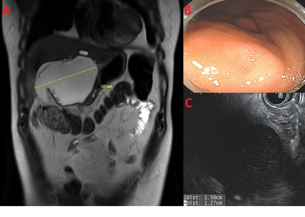
Figure: Ectopic pancreas with pseudocyst in the gastric antrum
The MRI abdomen demonstrated a large fluid collection inferior to the left lobe, measuring 12.7 x 7.5 x 7.7 cm, contiguous with EP, and consistent with pseudocyst (A). Endoscopic appearance of a subepithelial lesion found in the gastric antrum at the EP site (B). Sonographically, the EP appeared to originate from the submucosa, measuring 13 mm by 16 mm (C).
EP: ectopic pancreas
Disclosures: Hasan Saleh indicated no relevant financial relationships. Salvatore Crucillà indicated no relevant financial relationships. Jessica Petrov indicated no relevant financial relationships. Bhaumik Brahmbhatt indicated no relevant financial relationships. Michele Lewis indicated no relevant financial relationships.
Hasan Saleh, MD, MBA1, Salvatore Crucillà, 2, Jessica Petrov, MD, MSc1, Bhaumik Brahmbhatt, MBBS, MD1, Michele Lewis, MD1. P2336 - Ectopic Pancreas with Pseudocyst: An Elusive Cause of Recurrent Abdominal Pain, ACG 2025 Annual Scientific Meeting Abstracts. Phoenix, AZ: American College of Gastroenterology.