Abhi K. Singh, MD1, Daksesh B. Patel, DO2, Janis M. Atkinson, MD3 1St. Francis Hospital, Evanston, IL; 2St. Francis Hospital, Chicago, IL; 3Saint Francis Hospital Evanston, Wilmette, IL Introduction: Ciliated foregut cyst is a rare clinical entity and has been sporadically reported in literature. These relatively benign cysts result from abnormal development of the primitive foregut. When these cystic lesions arise from pancreas, their distinction from more common pancreatic cystic lesion solely based on imaging finding is extremely difficult. Hence, a histologic diagnosis must be pursued given the reports of malignant transformation and need for close follow up.
Case Description/
Methods: A 62-year-old white female was referred for evaluation of an incidentally detected cystic lesion in the pancreatic head. A 12 mm cystic lesion was initially detected during work up for hematuria in 2023. She denied history of chronic alcoholism, acute or chronic pancreatitis, gallstone or common bile duct (CBD) stone, and abdominal trauma. Follow up imaging a year later showed an increase in size to 20 mm without dilation of the main pancreatic duct. She remained asymptomatic. Her liver enzymes were normal and serum amylase was noted to be 28 IU/L (21-101 IU/L) while tumor markers were within the normal limits. [Carcinoembryonic antigen (CEA): 2.5 ng/ml and Carbohydrate antigen 19-9(CA 19-9): 28 U/ml]. Given progressive nature of the lesion, patient underwent an endoscopic ultrasound (EUS) in February, 2025 which showed a 15 mm x 10 mm anechoic lesion in the pancreatic head with two compartments without any septae or internal debris. About 3 ml of thick bloody fluid was aspirated and sent for cytology, amylase and CEA. No calcification, mass or lymphadenopathy were noted.The fine needle aspiration (FNA) cytology showed ciliated foregut epithelium. The cyst fluid amylase was noted to be 1214 IU/L while CEA was 7 ng/ml (0-5 ng/ml). She has a follow up MRI in 6 months to assess the progression of lesion.
Discussion: Ciliated foregut cyst of pancreas although rare, should be considered in the differential diagnosis of cystic lesion of pancreas. Despite the advances in the imaging technique, radiological diagnosis remains elusive.The use of EUS guided FNA has revolutionized the management of these cystic lesions. Obtaining an adequate cytology is paramount for distinguishing these benign cysts from those with malignant potential. Cystic fluid amylase level and tumor markers can serve as a valuable tool in the differential diagnosis of cystic lesion. However, caution should be exercised while interpreting pancreatic cyst fluid tumor markers as relying exclusively on these ancillary tests may lead to misdiagnosis.
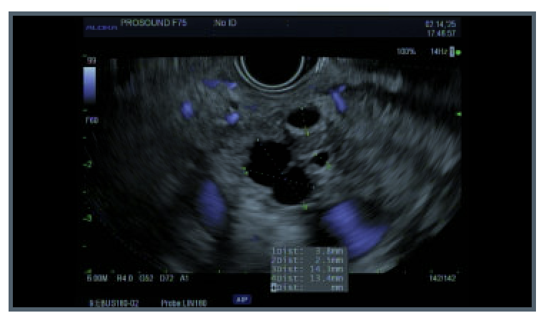
Figure: An endoscopic ultrasound showing 15 mm x 10 mm anechoic lesion in head of pancreas without septae or internal debris
Figure: Low resolution photograph (H & E stain x 200) showing a lining of benign, ciliated epithelial cells consistent with the diagnosis of foregut cyst.
Abhi K. Singh, MD1, Daksesh B. Patel, DO2, Janis M. Atkinson, MD3. P2330 - Ciliated Foregut Cyst of the Pancreas: A Rare Diagnostic Consideration, ACG 2025 Annual Scientific Meeting Abstracts. Phoenix, AZ: American College of Gastroenterology.