Jacob Lampenfeld, MD1, Corinne Zalomek, MD1, Husayn F. Ramji, MD2, Yucel Aydin, MD3, Kaitlin Occhipinti, MD3 1Tulane School of Medicine, New Orleans, LA; 2Tulane University School of Medicine, New Orleans, LA; 3Tulane University, New Orleans, LA Introduction: Gastrointestinal stromal tumors (GISTs) are a rare mesenchymal neoplasm of the gastrointestinal tract, most commonly arising in the stomach. Clinical presentations can vary widely, including incidental findings on imaging. This case highlights an atypical presentation of a large GIST initially misdiagnosed as pneumonia and pleural effusion.
Case Description/
Methods: A 60 year old male with a history of alcohol and substance abuse presented for progressive dyspnea, fatigue, enlarging left sided abdominal mass for three months, and recent development of melena. The patient had previously gone to two other hospitals for care, both of which he was diagnosed with pneumonia and pleural effusion.
He was hemodynamically stable, but appeared cachectic with a palpable left sided abdominal mass. Laboratory studies were significant for hemoglobin 4.5 g/dL. Contrasted computed tomography (CT) revealed a 24 cm intra-abdominal mass causing adjacent organs to shift, left hemidiaphragm elevation and mild compression of the left ventricle.
Upper endoscopy showed extrinsic compression of the gastric body and fundus; no intraluminal lesions or bleeding. Biopsy confirmed GIST. The patient was referred to Oncology for neoadjuvant chemotherapy followed by resection. Discussion: GIST most typically presents as abdominal pain, melena or hematemesis, fatigue, or unexplained weight loss; however, it can also be incidentally detected on imaging or endoscopy.
This patient’s initial presentation was dyspnea, presumed to be pulmonary in origin, and treated as such multiple times. It ultimately prompted further imaging to detect true etiology as a GIST causing diaphragmatic elevation and secondary cardiopulmonary effects.
This case highlights the importance of maintaining a broad differential diagnosis for common complaints. Further, it reinforces the value of reevaluating initial diagnoses when standard therapies fail to improve clinical status.
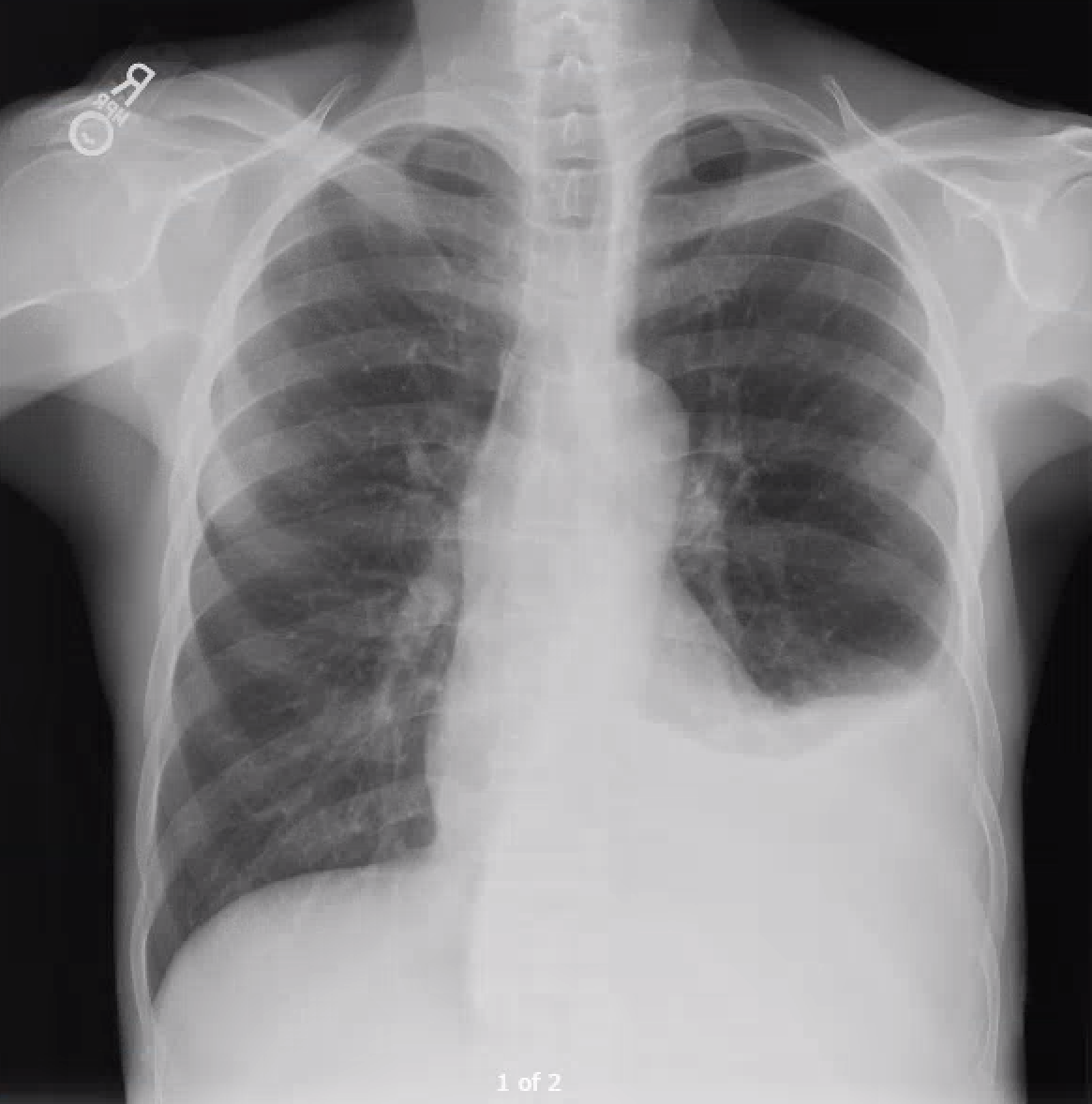
Figure: Chest X-ray at admission showing left costophrenic opacity
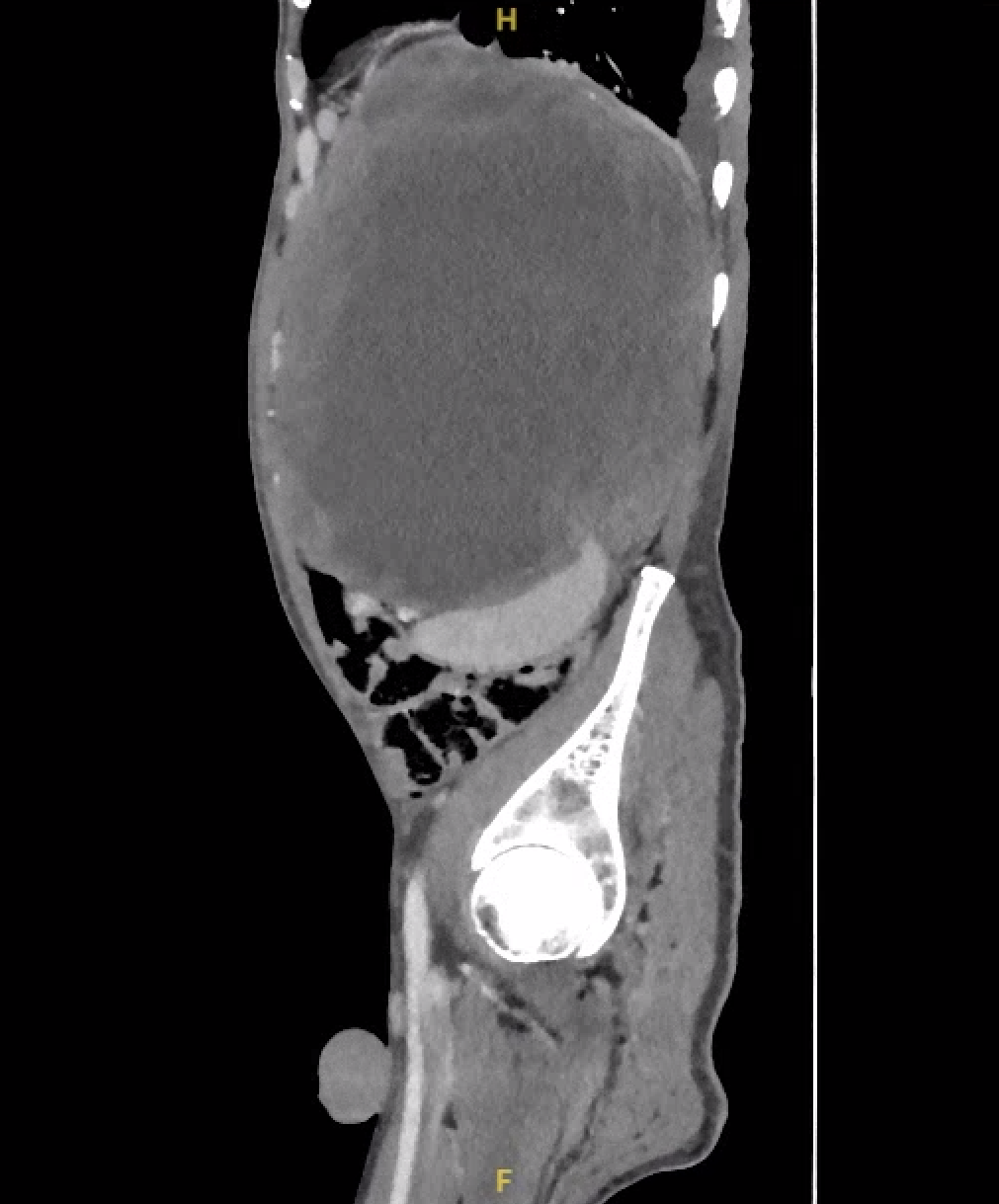
Figure: CT of the abdomen & pelvis (lateral view) showing large intra-abdominal mass causing hemidiaphragm elevation
Disclosures: Jacob Lampenfeld indicated no relevant financial relationships. Corinne Zalomek indicated no relevant financial relationships. Husayn Ramji indicated no relevant financial relationships. Yucel Aydin indicated no relevant financial relationships. Kaitlin Occhipinti indicated no relevant financial relationships.
Jacob Lampenfeld, MD1, Corinne Zalomek, MD1, Husayn F. Ramji, MD2, Yucel Aydin, MD3, Kaitlin Occhipinti, MD3. P2122 - Massive GIST Masquerading as a Pleural Effusion: A Case Report, ACG 2025 Annual Scientific Meeting Abstracts. Phoenix, AZ: American College of Gastroenterology.