Patrick Yang, MD, Sarah Rahni, DO, Menasche Krupka, MD, Cynthia Cohen, MD, Shireen Pais, MD Westchester Medical Center, Valhalla, NY Introduction: Gastric squamous cell cancer (GSCC) is a rare histologic variant of gastric cancer. While over 90% of gastric cancers are adenocarcinoma, only 0.040-0.5% are squamous cell cancer. The clinical manifestations are often nonspecific and similar to other gastric cancers. Here, we report a case of GSCC with late presentation.
Case Description/
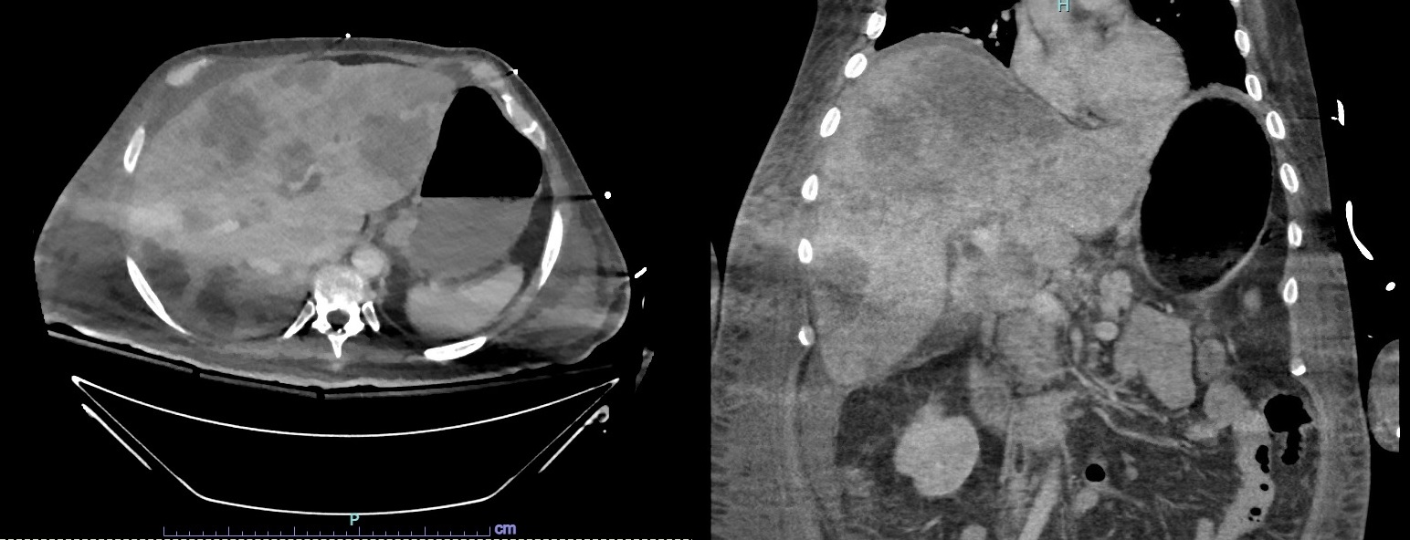
Methods: 52-year-old male smoker with 36 pack years, no prior cancer history, but family history of gastric cancer in his father at age 59, presented with lethargy and melena after recent admission a week prior for pulmonary emboli. Previous admission was prompted by night sweats, decreased appetite, jaundice, and 40 pound weight loss. CT abdomen at that time revealed innumerable hepatic lesions with peri-aortic lymphadenopathy concerning for metastatic disease. Biopsy of liver lesions was consistent with poorly differentiated squamous cell carcinoma, staining positive for p40 and CK5/6 but negative for S100, TTF1 and NKX3.1.
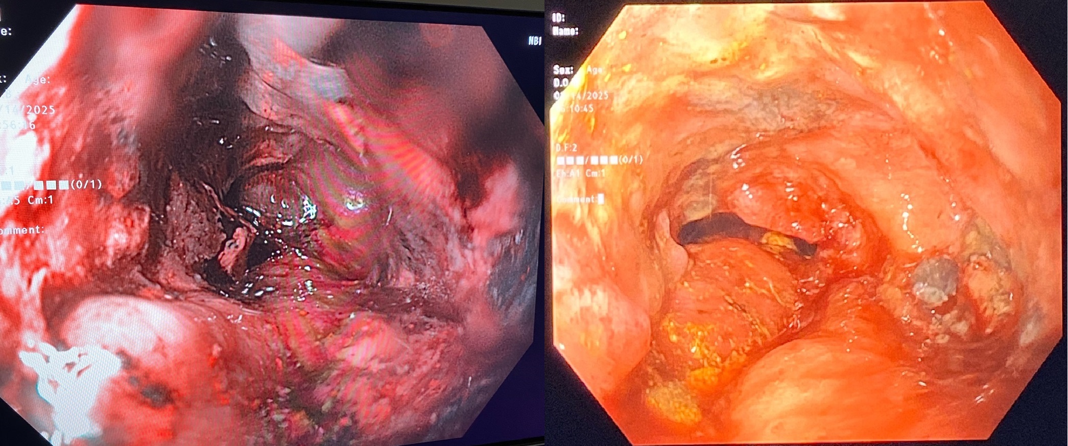
Patient was admitted to the ICU with hypotension requiring vasopressors and concern for septic shock. CEA level was elevated to 80.6 ng/ml and AFP was normal at 0.37 ng/ml. EGD revealed a circumferential gastric mass over 10cm in length with involvement of the proximal body and fundus. Coffee ground gastric contents without fresh blood was visualized. Biopsy of the gastric mass was positive for p40 and consistent with keratinizing squamous cell carcinoma, with ulceration of the mucosa, marked inflammation and fungal infiltration. Imaging for full metastatic work-up did not reveal spread outside of the liver and periaortic lymph nodes. He was critically ill with shock, multiorgan failure, acidosis, and encephalopathy in the setting of newly diagnosed metastatic gastric cancer. Despite broad spectrum coverage including micafungin, he had a rapid decline and ultimately passed away after 3 days. Discussion: GSCC has poor prognosis as it is often diagnosed at an advanced stage with frequent lymphovascular serosal involvement. Typically, it is more prevalent in men, past the 6th decade of life, with positive smoking history, which matches this patient’s demographic. While the pathogenesis of GSCC remains unclear, proposed hypotheses involve squamous cell metaplasia or nests of ectopic squamous epithelium developing into SCC in the setting of chronic inflammation or viral infection. There is no consensus regarding optimal treatment strategy. Development of earlier detection methods would be pivotal towards improving prognosis.
Figure: Large, erythematous, friable 10cm gastric mass with areas of necrosis and ulceration involving the proximal body and fundus. Multiple cold forceps biopsies obtained.
Figure: CT Abdomen with contrast: Axial section shown on left and coronal section on the right. There are innumerable hypodense masses throughout the liver concerning for metastatic disease. No significant abnormalities in the stomach appreciated on CT scan.
Disclosures: Patrick Yang indicated no relevant financial relationships. Sarah Rahni indicated no relevant financial relationships. Menasche Krupka indicated no relevant financial relationships. Cynthia Cohen indicated no relevant financial relationships. Shireen Pais indicated no relevant financial relationships.
Patrick Yang, MD, Sarah Rahni, DO, Menasche Krupka, MD, Cynthia Cohen, MD, Shireen Pais, MD. P2113 - A Rare Case of Gastric Squamous Cell Carcinoma, ACG 2025 Annual Scientific Meeting Abstracts. Phoenix, AZ: American College of Gastroenterology.