University of California San Francisco (Fresno) Fresno, CA
Katherine Jones, DO1, Arpine Petrosyan, MD1, Rajarajeshwari Ramachandran, MD2 1University of California San Francisco (Fresno), Fresno, CA; 2Fresno Veterans Affairs Medical Center, Fresno, CA Introduction: Tumor Melanosis (TM) is a rare manifestation found at the site of completely regressed melanoma. Although these lesions have a phenotypic appearance of melanoma, biopsies will demonstrate aggregates of melanin-laden macrophages in the absence of malignant melanocytes (1-3). Few cases of TM have been described in the literature to date. Here we present a rare case of a 75-year-old male with TM following treatment of gastric melanoma with pembrolizumab.
Case Description/
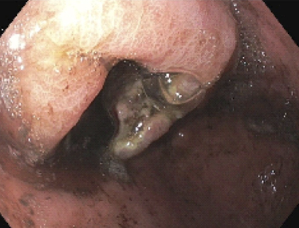
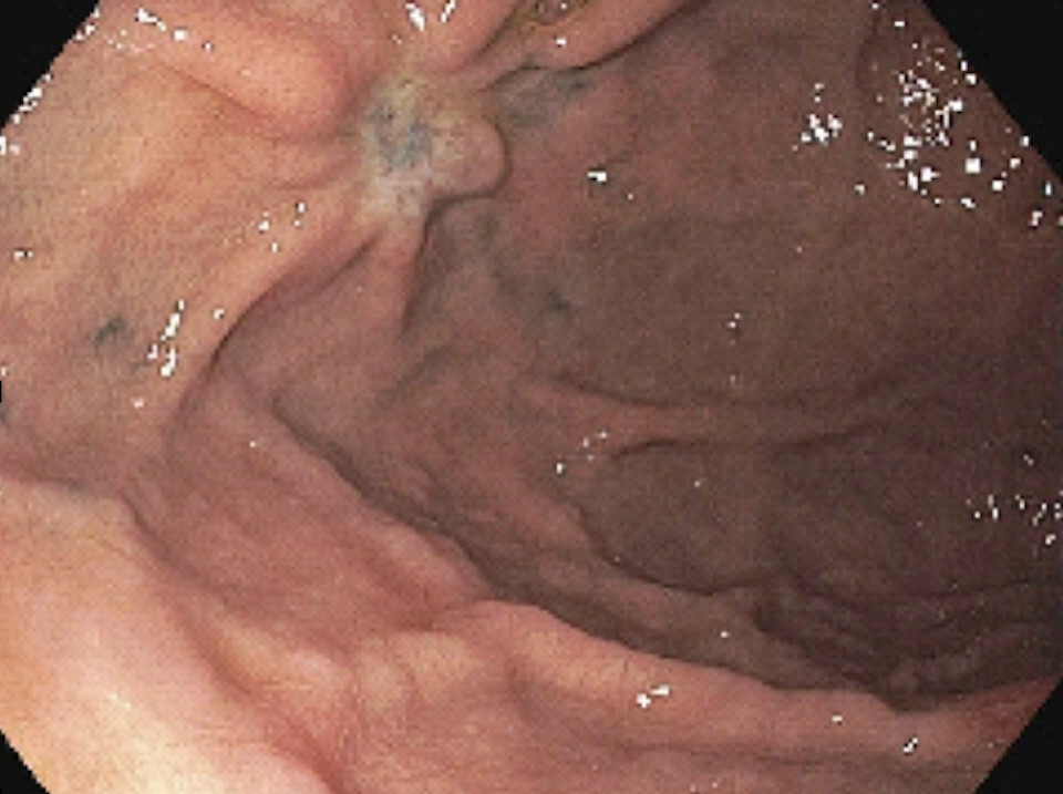
Methods: A 75-year-old male presented to the hospital for evaluation of melena and acute anemia. Esophagogastroduodenoscopy (EGD) demonstrated multiple darkly pigmented ulcerated gastric masses (largest measuring 3-4 cm) in the body and fundus. Biopsies revealed an epithelioid neoplasm in the submucosa with intracytoplasmic pigment, suspicious for melanin. Immunohistochemical staining confirmed Melan-A, SOX10, and S100, consistent with the diagnosis of gastric melanoma. After completion of pembrolizumab therapy, repeat EGD was performed and a 3 mm pigmented lesion was seen in the gastric fundus (corresponding to one of the sites of prior gastric melanoma), surrounded by a few pigmented satellite lesions 1-2 mm in size. Biopsies of these lesions were positive for Fontana-Masson stain, suggesting a melanin-like pigment, as well as CD68 confirming the presence of macrophages. Melan-A was negative, and this excluded the possibility of residual melanoma. Overall, the histologic findings were consistent with TM. Discussion: TM is a rare condition and most cases reported in the literature involve the skin. Our case will add to the limited number of cases of gastric TM reported to date (4). Histopathological examination is crucial in differentiating residual melanoma from TM.
References:
Bari O, Cohen PR. Tumoral Melanosis Associated with Pembrolizumab-Treated Metastatic Melanoma. Cureus. 2017;9(2):e1026. Published 2017 Feb 13. doi:10.7759/cureus.1026
Burzi L, Alessandrini AM, Quaglino P, Piraccini BM, Dika E, Ribero S. Cutaneous Events Associated with Immunotherapy of Melanoma: A Review. J Clin Med. 2021;10(14):3047. Published 2021 Jul 8. doi:10.3390/jcm10143047
Staser K, Chen D, Solus J, et al. Extensive tumoral melanosis associated with ipilimumab-treated melanoma. Br J Dermatol. 2016;175(2):391-393. doi:10.1111/bjd.14474
Bronswijk M, Sagaert X, Bechter OE. Gastric Tumoral Melanosis: a Rare Manifestation of Successfully Treated Malignant Melanoma. Clin Gastroenterol Hepatol. 2020;18(8):A37-A38. doi:10.1016/j.cgh.2019.02.006
Figure: Figure 1A: EGD image at initial diagnosis of melanoma showing a pigmented gastric mass with ulceration
Figure: Figure 1B: EGD image following treatment with pembrolizumab showing 3 mm pigmented lesion in gastric fundus
Disclosures: Katherine Jones indicated no relevant financial relationships. Arpine Petrosyan indicated no relevant financial relationships. Rajarajeshwari Ramachandran indicated no relevant financial relationships.
Katherine Jones, DO1, Arpine Petrosyan, MD1, Rajarajeshwari Ramachandran, MD2. P2107 - Tumor Melanosis Following Treatment of Gastric Melanoma With Pembrolizumab, ACG 2025 Annual Scientific Meeting Abstracts. Phoenix, AZ: American College of Gastroenterology.