Alexander Siegel, MD1, Clive Miranda, DO2, Mohammed Qasswal, MD3, Robert Kizer, MD3 1CHI Health Creighton University Medical Center, Elkhorn, NE; 2CHI Health Creighton University Medical Center, Omaha, NE; 3Creighton University Medical Center, Omaha, NE Introduction: Breast cancer is the most common form of cancer diagnosed in women worldwide, with an estimated mortality rate of 6.9%. Breast cancer is typically detected through routine screening mammography, which is effective in identifying early-stage disease. However, certain subtypes may present atypically. Although well documented, gastric metastasis from primary breast cancer is rare, only occurring in about 0.3% of cases.
Case Description/
Methods: A 44-year-old Asian female-immigrant with a past medical history significant for gastroesophageal reflux disease, anemia and latent tuberculosis presented with a three-day history of increasing abdominal pain. Four days earlier, she had presented to an outside hospital with similar symptoms but left against medical advice. Abdominal CT performed at the outside hospital revealed gastric wall thickening with scattered nodules in the bladder, ureters, vulva and perineum, concerning for metastatic disease. Due to concern for a primary gastrointestinal malignancy, gastroenterology was consulted for further evaluation.
Upper and lower endoscopies revealed multiple submucosal nodules in the stomach, duodenum, and the entire colon. Histopathologic analysis of the gastric and colonic biopsies demonstrated estrogen receptor and progesterone receptor positive, HER2-negative breast cancer. The patient had no family history of breast cancer and underwent screening mammography three months prior to presentation where no mass was identified. Evan after the diagnosis of metastatic breast cancer was confirmed, whole-body CT/PET imaging revealed no discreate breast mass. The patient was started on leuprolide, and outpatient follow-up was arranged with oncology to initiate treatment with ribociclib. Discussion: Screening mammography is the gold standard for breast cancer surveillance and has been highly effective in detecting early-stage disease, particularly in patients without significant risk factors. With routine imaging every one to two years, over 90% of breast cancers are diagnosed before development of distant metastases. However, this approach may fail to detect rapidly progressive or non-mass-forming subtypes. This case underscores the diagnostic challenge when faced with atypical breast cancer presentations and emphasizes the importance of including breast cancer in the differential when radiographic and endoscopic evaluation is consistent with metastatic disease.
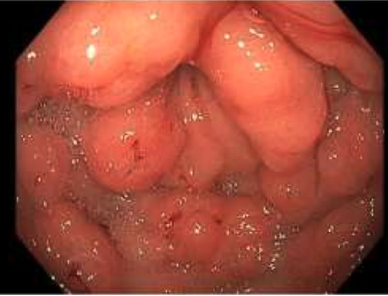
Figure: Endoscopic image of nodules located at the pylorus
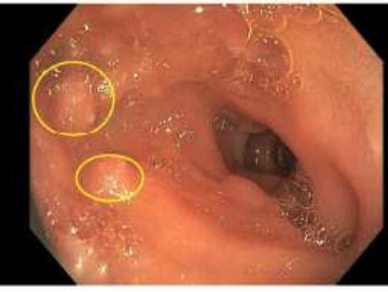
Figure: Endoscopic image of nodules located in the duodenal bulb
Disclosures: Alexander Siegel indicated no relevant financial relationships. Clive Miranda indicated no relevant financial relationships. Mohammed Qasswal indicated no relevant financial relationships. Robert Kizer indicated no relevant financial relationships.
Alexander Siegel, MD1, Clive Miranda, DO2, Mohammed Qasswal, MD3, Robert Kizer, MD3. P2077 - Atypical Presentation of Metastatic Breast Cancer in a 44-Year-Old Female: A Case Report, ACG 2025 Annual Scientific Meeting Abstracts. Phoenix, AZ: American College of Gastroenterology.