Ahmed Mohamed Ebeid, MD1, Nakul Ganju, MD1, Alaa Abdelsamad, MD1, Linda Uzo. Amara, MD1, Juan C. Santiago-Gonzalez, MD, MSc1, Mesay Asfaw, MD2, Amro Abdellatief, MD1, Maryam Foroughi, MD1, Angesom Kibreab, MD1 1Howard University Hospital, Washington, DC; 2Howard University, Washington, DC Introduction: Gastric adenocarcinoma commonly metastasizes to the liver, peritoneum, and lungs, with bone involvement being rare (0.9%–13.4% of cases). Initial presentation through isolated osteolytic spinal lesions, without gastrointestinal symptoms, is even more uncommon. This atypical presentation and low suspicion for a gastrointestinal primary complicate the diagnosis. We report a rare case of gastric adenocarcinoma identified through vertebral osteolytic lesions, underscoring the need to consider gastrointestinal malignancies in patients with unexplained bone disease
Case Description/
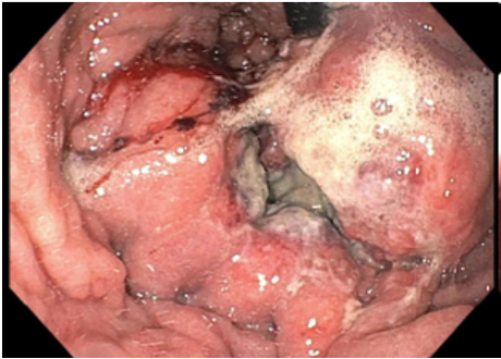
Methods: A 66-year male patient with history of acid reflux presented with generalized fatigue, loss of appetite with significant weight loss, and worsening back pain for 6 months. Physical exam revealed point tenderness over the thoracic and lumbar spine. Labs revealed a hemoglobin of 9.8 gm/dL, Mean Corpuscular Volume: 72.3 L, Alkaline Phosphatase: 188 U/L, Ca 10.7, Intrinsic Factor Blocking AB: Pos, CEA: 7.49, CA 19-9: normal. CT abdomen/pelvis revealed multiple lytic lesions throughout the osseous structures of T0, L3-L5, lesions adjacent to the splenic hilum and greater curvature of the stomach and extensive retroperitoneal celiac lymphadenopathy. Upper endoscopy revealed a large fungating, infiltrative and ulcerated mass without bleeding involving the cardia, fundus and the greater curvature of the stomach (Figure 1). Biopsy revealed inflamed tissue with infiltration by poorly differentiated malignant cells highlighted by CK20 and focally positive for CDX2. IR CT-guided core needle aspiration and biopsy of the spinal lesion revealed metastatic epithelial cells with glandular architectures and with moderate to high grade nuclear features (Figure 2) Discussion: Intestinal-type gastric adenocarcinoma, characterized by co-expression of CK20 and CDX2, is a major histologic subtype of gastric cancer. It commonly affects older adults and males, typically arising in the distal stomach, including the body and antrum. This subtype is frequently associated with chronic gastritis and intestinal metaplasia, often triggered by Helicobacter pylori
infection. Although this cancer type usually has a favorable prognosis and infrequently metastasizes, our patient’s diagnosis of a poorly differentiated tumor and his sole presenting symptom of skeletal pain, without gastrointestinal complaints, reflect a more aggressive clinical course and underscore the diagnostic challenges it can pose
Figure: Figure 1: An esophagogastroduodenoscopy revealing: a large fungating, infiltrative and ulcerated mass without bleeding involving the cardia, fundus and the greater curvature of the stomach
Figure: Figure 2: IR CT-guided core needle aspiration and biopsy of the spinal lesion revealed metastatic epithelial cells with glandular architectures and with moderate to high grade nuclear features
Disclosures: Ahmed Mohamed Ebeid indicated no relevant financial relationships. Nakul Ganju indicated no relevant financial relationships. Alaa Abdelsamad indicated no relevant financial relationships. Linda Amara indicated no relevant financial relationships. Juan Santiago-Gonzalez indicated no relevant financial relationships. Mesay Asfaw indicated no relevant financial relationships. Amro Abdellatief indicated no relevant financial relationships. Maryam Foroughi indicated no relevant financial relationships. Angesom Kibreab indicated no relevant financial relationships.
Ahmed Mohamed Ebeid, MD1, Nakul Ganju, MD1, Alaa Abdelsamad, MD1, Linda Uzo. Amara, MD1, Juan C. Santiago-Gonzalez, MD, MSc1, Mesay Asfaw, MD2, Amro Abdellatief, MD1, Maryam Foroughi, MD1, Angesom Kibreab, MD1. P2071 - The Spine as a Clue: Unmasking Gastric Adenocarcinoma Through Bone Lesions, a Rare Case Report, ACG 2025 Annual Scientific Meeting Abstracts. Phoenix, AZ: American College of Gastroenterology.