Essam Rashad, MBBCh1, Umer Farooq, MD2, Abdul Haseeb, MD, MPH3 1Parkview Medical Center, Fort Wayne, IN; 2Saint Louis University School of Medicine, St. Louis, MO; 3Midwest Digestive Health & Nutrition, Hinsdale, IL Introduction: Gastric involvement in AL amyloidosis occurs in fewer than 20% of patients. Endoscopic findings are often nonspecific, and normal findings do not exclude disease. Amyloid may deposit in different layers of the gastrointestinal tract, which can pose a diagnostic challenge. Findings include nodules, erythema, ulceration, mucosal friability or thickening, and normal mucosa. We present this case to highlight delayed presentation of gastric amyloidosis manifested as gastroparesis and nodular lesions seen on endoscopy.
Case Description/
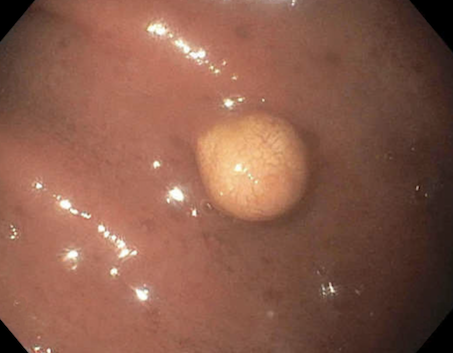
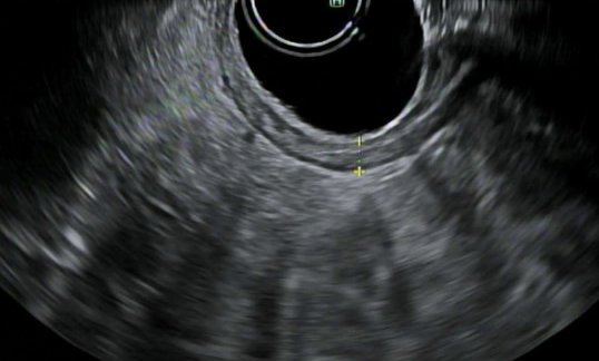
Methods: A 50-year-old non-diabetic woman with a history of multiple myeloma complicated by AL (amyloid light chain) amyloidosis in 2013 and renal transplantation in 2021 on immunosuppressive therapy initially presented with epigastric pain, nausea, and early satiety. Initial laboratory evaluation was unremarkable, and esophagogastroduodenoscopy (EGD) demonstrated mildly erythematous gastric mucosa without biopsy sampling. She was treated empirically with a short course of proton pump inhibitors; however, persistent symptoms and a 6.3 kg weight loss over the subsequent 10 months prompted further evaluation, including a gastric emptying study consistent with grade 4 gastroparesis and a repeat EGD revealing multiple 2–3 mm non-bleeding nodules in the gastric body and antrum. Endoscopic ultrasound (EUS) demonstrated normal gastric wall thickness. Biopsies obtained from the gastric wall, antrum, and nodule were negative for Helicobacter pylori but were universally positive for deposition on Congo red staining. Patient was managed symptomatically with antiemetics, gabapentin, and analgesia and referred to a tertiary center to discuss treatment. Discussion: Gastric involvement in AL amyloidosis typically manifests with non-specific symptoms such as weight loss, abdominal pain, and nausea or vomiting. Endoscopic findings are similarly variable, ranging from normal-appearing mucosa to erythema, erosions, nodularity, and subepithelial lesions (SELs). In our patient, gastroparesis was likely attributable to neuromuscular infiltration by AL amyloid, in addition to the presence of nodular lesions. A high index of clinical suspicion should be maintained in patients with known amyloidosis who present with non-specific upper gastrointestinal symptoms.
Figure: Single 2 mm mucosal papule in the gastric antrum representing gastric amyloid.
Figure: EUS showing normal gastric wall thickness.
Disclosures: Essam Rashad indicated no relevant financial relationships. Umer Farooq indicated no relevant financial relationships. Abdul Haseeb indicated no relevant financial relationships.
Essam Rashad, MBBCh1, Umer Farooq, MD2, Abdul Haseeb, MD, MPH3. P2065 - Atypical Gastrointestinal Manifestations of AL Amyloidosis: A Case of Gastroparesis and Nodular Gastric Lesions, ACG 2025 Annual Scientific Meeting Abstracts. Phoenix, AZ: American College of Gastroenterology.