Oghuz Anwar, DO1, Moiz Shah, MBChB1, Jeffrey Laczek, MD2 1Kaiser Permanente, Gaithersburg, MD; 2Kaiser Permanente, Upper Marlboro, MD Introduction: Gastric adenocarcinoma typically recurs as solid masses in the peritoneum, lymph nodes, or liver. While cystic transformation has been reported in other malignancies—such as pancreatic, renal, and thymic tumors—it is not well described in gastric cancer. Carcinoembryonic antigen (CEA) is a well-established tumor marker in gastrointestinal cancers and is used in pancreatic cyst fluid to distinguish mucinous from non-mucinous lesions, but its role in other cystic lesions is unclear. We present a case of gastric adenocarcinoma treated with chemotherapy and surgery, later found to have a large cystic recurrence with markedly elevated cyst fluid CEA, raising awareness of this atypical recurrence pattern and the potential diagnostic utility of CEA in cystic fluid analysis.
Case Description/
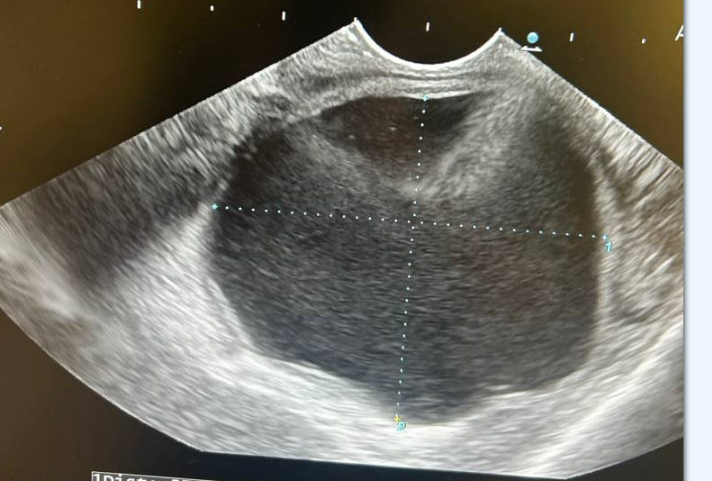
Methods: A 52-year-old man with a history of gastroesophageal reflux disease presented with dyspepsia and a 30-lb weight loss. Workup revealed HER2-negative, mismatch repair (MMR)-intact gastric adenocarcinoma (T3N1) spanning the GE junction to the antrum. He underwent neoadjuvant chemotherapy followed by total gastrectomy with complete pathologic response (ypT0N0). Patient received adjuvant chemotherapy, and initial surveillance imaging showed no recurrence. Four months later, CT revealed new hypodense, cystic-appearing lesions near the gastric reconstruction. Positron emission tomography/CT showed no fluorodeoxyglucose (FDG) uptake in these lesions. EUS revealed a 58 x 51 mm cystic collection; 96 mL of orange fluid was aspirated. Cytology confirmed carcinoma consistent with gastric origin, and fluid analysis showed a markedly elevated CEA level of 2,907 ng/mL. Discussion: This case illustrates an unusual cystic recurrence following curative-intent treatment of gastric adenocarcinoma. CEA is traditionally used to distinguish mucinous from non-mucinous pancreatic cysts. Its elevated concentration in cyst fluid here perhaps suggests a broader diagnostic role. Elevated cyst fluid CEA can raise suspicion for malignant recurrence in patients with prior GI malignancy—even in the setting of complete pathologic response and non-FDG-avid lesions. When cytology is diagnostic, as it was here, CEA may serve as a useful confirmatory marker. Conversely, in cases where cytologic findings are inconclusive, cyst fluid CEA may serve as a valuable diagnostic “double check” to support clinical decision-making. Ultimately, elevated cyst fluid CEA may be a valuable adjunctive marker for malignant recurrence and warrants further study.
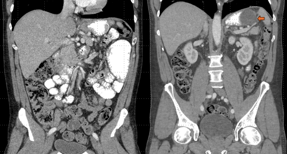
Figure: Surveillance CT with stable post-surgical changes and no evidence of metastatic disease (Left). Follow up imaging four months later with new hypodense, cystic appearing lesions adjacent to the gastric reconstruction, largest measuring 5.7 cm (Right).
Figure: Cystic fluid collection seen on endoscopic ultrasound before aspiration
Disclosures: Oghuz Anwar indicated no relevant financial relationships. Moiz Shah indicated no relevant financial relationships. Jeffrey Laczek indicated no relevant financial relationships.
Oghuz Anwar, DO1, Moiz Shah, MBChB1, Jeffrey Laczek, MD2. P2064 - Cystic Recurrence of Gastric Adenocarcinoma with Elevated Cyst Fluid CEA: A Rare Occurrence and a Potentially Useful Marker, ACG 2025 Annual Scientific Meeting Abstracts. Phoenix, AZ: American College of Gastroenterology.