University of Medicine and Health Sciences Clinton Township, MI
Jeffrey Sharza, BS1, Manvith Munagala, MD2, Michel Sabbagh, MD2, Nathalia Millan-Borrero, MD2, Georgette Cooke, DO2 1University of Medicine and Health Sciences, Clinton Township, MI; 2McLaren Macomb Hospital, Mount Clemons, MI Introduction: Splenic autoinfarction is the primary cause of autosplenectomy in patients with sickle cell disease. Patients with the more mild sickle cell trait can be prone to recurrent infarction of the spleen with significant oxygen changes most commonly seen in drastic elevation changes such as frequent skydiving or scuba diving. This is likely due to changes in the oxygen dissociation curve at different elevations triggering a transient hypercoagulable state. Other and more common causes of splenic infarcts are autoimmune-mediated including lupus and antiphospholipid syndrome. This case is significant because in the absence of common risk factors such as barometric changes and diagnosed autoimmune conditions there is a high suspicion of a sepsis-induced splenic infarct.
Case Description/
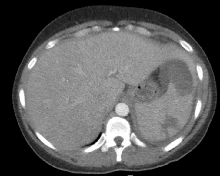
Methods: A 40 year old woman with a past medical history of sickle cell trait, hypothyroidism post-thyroidectomy, uterine fibroids and iron deficiency anemia presented to the emergency room for a week of increasing diffuse abdominal pain, hot flashes, night sweats, cold clammy skin, nausea, vomiting and 3-day history of intermittent cough. She also endorsed recent dark stools but has been on longstanding supplemental iron therapy. A couple weeks prior, she had a uterine biopsy for her fibroids and was scheduled for a total hysterectomy. Initial vitals in the emergency department revealed a heart rate of 139 bpm and fever of 37.9°C. Labs were significant for leukocytosis of 33k WBC/µL, hemoglobin of 7.4 g/dL and a lactate of 2.4 mmol/L consistent with sepsis. Blood cultures and urine culture grew Streptococcus pyogenes. Hemoglobin electrophoresis showed 30% HbS. CT abdomen and pelvis demonstrated wedge hypodensities in the spleen consistent with infarcts rarely seen in patients with sickle cell trait. Per her history, she has not recently travelled, flown, skydived or scuba dove. Further workup for hypercoagulability revealed elevated LDH and fibrinogen, and negative lupus anticoagulant, anticardiolipin and beta-2 glycoprotein. Discussion: This case presents a splenic infarct in a patient with sickle cell trait without common triggers. While SCT-associated infarcts usually result from hypoxia, this instance points to sepsis-induced coagulopathy as the likely cause. The systemic inflammation and microvascular thrombosis during sepsis may have created a "second hit," inducing sickling and splenic vaso-occlusion in this ‘higher-risk’ patient. This case may broaden our understanding of splenic infarcts in SCT.
Figure: The spleen shows multiple wedge-shaped hypodensities, which are suggestive of splenic infarcts.
Disclosures: Jeffrey Sharza indicated no relevant financial relationships. Manvith Munagala indicated no relevant financial relationships. Michel Sabbagh indicated no relevant financial relationships. Nathalia Millan-Borrero indicated no relevant financial relationships. Georgette Cooke indicated no relevant financial relationships.
Jeffrey Sharza, BS1, Manvith Munagala, MD2, Michel Sabbagh, MD2, Nathalia Millan-Borrero, MD2, Georgette Cooke, DO2. P2062 - Atypical Splenic Infarct in Patient With Sickle Cell Trait, ACG 2025 Annual Scientific Meeting Abstracts. Phoenix, AZ: American College of Gastroenterology.