Anmol Mittal, MD, MBA, Ahmed Al-Khazraji, MD Rutgers New Jersey Medical School, Newark, NJ Introduction: Gastric amyloidosis is a rare and often underrecognized manifestation of systemic amyloid deposition, characterized by the abnormal accumulation of amyloid proteins within the gastric mucosa. Its clinical presentation can be variable, ranging from nonspecific gastrointestinal symptoms to overt bleeding, making diagnosis challenging. Endoscopic evaluation plays a pivotal role in identifying characteristic mucosal changes, which, when coupled with histopathological confirmation, can establish the diagnosis. This case highlights the endoscopic features and diagnostic process of gastric amyloidosis in a patient presenting with gastrointestinal bleeding.
Case Description/
Methods: A 70-year-old male with a medical history of hypertension, dyslipidemia, diabetes mellitus, and atrial fibrillation on Apixaban presented with melena and worsening shortness of breath. He was diagnosed with acute pulmonary edema, complicated by heart failure with reduced ejection fraction, necessitating ICU admission. The patient had undergone an endoscopic evaluation one month earlier for a suspected upper gastrointestinal bleed and was diagnosed with diffuse atypical angioectasia, treated with incomplete Argon Plasma Coagulation (APC).
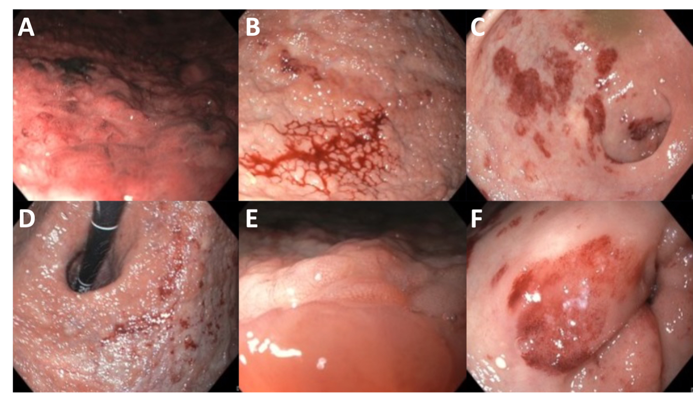
On repeat endoscopy, the following findings were observed:
1. Patchy, rose-colored gastritis with oozing in the antrum and a tendency to bleed.
2. Pit pattern mucosa visualized using Narrow Band Imaging (NBI).
3. Nodular raised gastric lesions with a pale, greyish background appearance in the body, fundus, and cardia, suggestive of mucosal ischemia from large protein/amyloid deposition. These findings are classic for gastric amyloidosis.
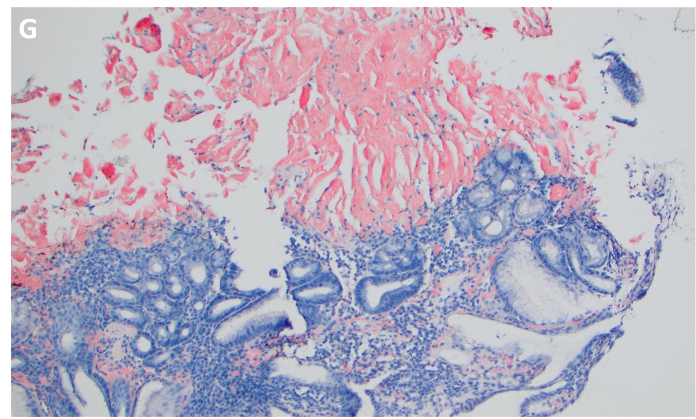
The diagnosis was confirmed through gastric biopsies, which showed positive Congo red staining, confirming the presence of gastric amyloidosis. Discussion: This case underscores the importance of considering gastric amyloidosis in patients with unexplained gastric mucosal abnormalities and bleeding, especially in those with complex medical histories. Endoscopic findings such as patchy gastritis, nodular lesions, and mucosal ischemia, along with advanced imaging techniques like Narrow Band Imaging, can provide critical clues. Confirmatory biopsy with Congo red staining remains the definitive diagnostic modality. Recognizing these features can facilitate timely diagnosis and appropriate management, potentially improving outcomes in affected patients.
Figure: Endoscopic images of upper endoscopy.
Figure: Histology slide with Congo Red staining.
Disclosures: Anmol Mittal indicated no relevant financial relationships. Ahmed Al-Khazraji indicated no relevant financial relationships.
Anmol Mittal, MD, MBA, Ahmed Al-Khazraji, MD. P2060 - Classic Endoscopic Features of Gastric Amyloidosis, ACG 2025 Annual Scientific Meeting Abstracts. Phoenix, AZ: American College of Gastroenterology.