David Guevara-Lazo, MD, Jorge D.. Machicado, MD, MPH University of Michigan, Ann Arbor, MI Introduction: A diffuse dilated pancreatic duct can be caused by different conditions, including pancreatic cancer, chronic pancreatitis and main duct intraductal papillary mucinous neoplasm (IPMN). Herein, we describe a case of a rare cause of dilation of the pancreatic duct.
Case Description/
Methods: A 69-year-old woman with type 2 diabetes mellitus presented with persistent diarrhea. After exhaustive work-up, this was determined to be secondary to metformin. Her workup included a CT of the abdomen that showed pancreatic atrophy and ductal dilation. A subsequent MRCP showed pancreas divisum, diffuse main duct dilation, and a 7×10×11 mm cystic lesion in the minor papilla suspicious for a santorinicele. An endoscopic ultrasound was done that showed no pancreatic malignancy, and no sampling was performed of the cystic lesion. The patient was followed with cross-sectional images, and eventually a MRCP done 2 years later showed enlargement of the santorinicele up to 25 mm and main duct dilation of 9 mm. Given the suspicion for an IPMN, a repeated EUS was performed, showing a cyst measuring 19x18 mm in the minor papilla with ductal dilation up to 7.1 mm in the head. Fine needle aspiration of the cystic lesion showed a low CEA (3.4 ng/mL) and cytology negative for malignancy. ERCP was later performed that confirmed the cystic lesion in the minor papilla communicating with the main pancreatic duct and consistent with a santorinicele. This was treated with a minor papilla sphincterotomy. In addition, a pancreatoscopy was performed that demonstrated a normal pancreatic duct epithelium. On follow-up MRCP a year later, the cystic lesion was no longer visible. Discussion: A santorinicele is a focal cystic dilatation of the terminal dorsal pancreatic duct and is often associated with ductal anomalies (e.g., pancreas divisum). It is thought to result from increased intraductal pressure due to relative stenosis at the minor papilla. Endoscopic interventions such as sphincterotomy can relieve ductal obstruction and symptoms. Given the cystic appearance, IPMN must be ruled out due to its malignant potential. In this case, the EUS findings and negative cytology excluded malignancies and guided therapeutic intervention.
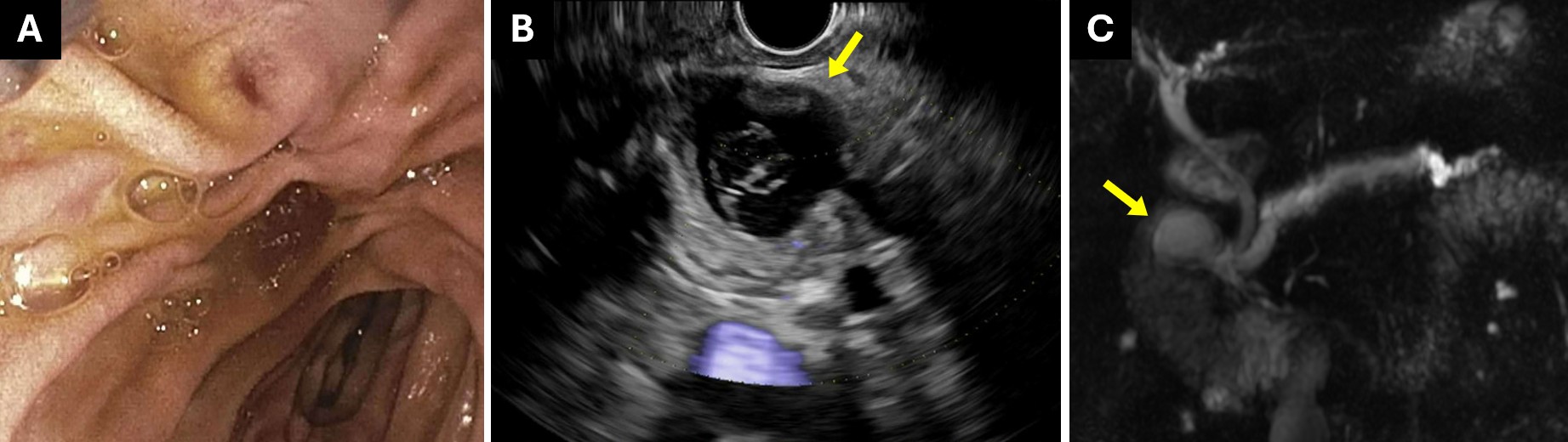
Figure: A) ERCP showing no morphological abnormalities of the minor papilla. B) EUS demonstrating a 19×18 mm cystic lesion at the level of the minor papilla. C) MRCP revealing a dilated pancreatic duct and a santorinicele.
Disclosures: David Guevara-Lazo indicated no relevant financial relationships. Jorge Machicado indicated no relevant financial relationships.
David Guevara-Lazo, MD, Jorge D.. Machicado, MD, MPH. P2354 - Santorinicele: A Rare Cause of Pancreatic Duct Dilation, ACG 2025 Annual Scientific Meeting Abstracts. Phoenix, AZ: American College of Gastroenterology.

 photo")