Fady Gendy, DO1, Michael Makar, MD1, Veneeth Antony, DO1, Aws Alameri, MD1, Patrick Dorion, MD1, Bradley D. Confer, DO1, Harshit S. Khara, MD2 1Geisinger Health System, Danville, PA; 2Geisinger Health System, Danville, NJ Introduction: Ectopic pancreatic tissue is defined as pancreatic tissue lacking any vascular or anatomic communication with the normal body of the pancreas. It is most commonly found in the foregut, specifically the stomach. While typically asymptomatic, it may cause symptoms when complications like inflammation, obstruction, or malignancy occur. In this report, we highlight a rare case of biliary obstruction caused by benign pancreatic acinar tissue, causing obstructive jaundice.
Case Description/
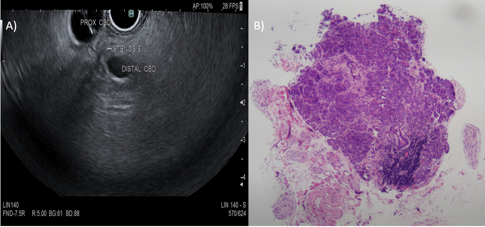
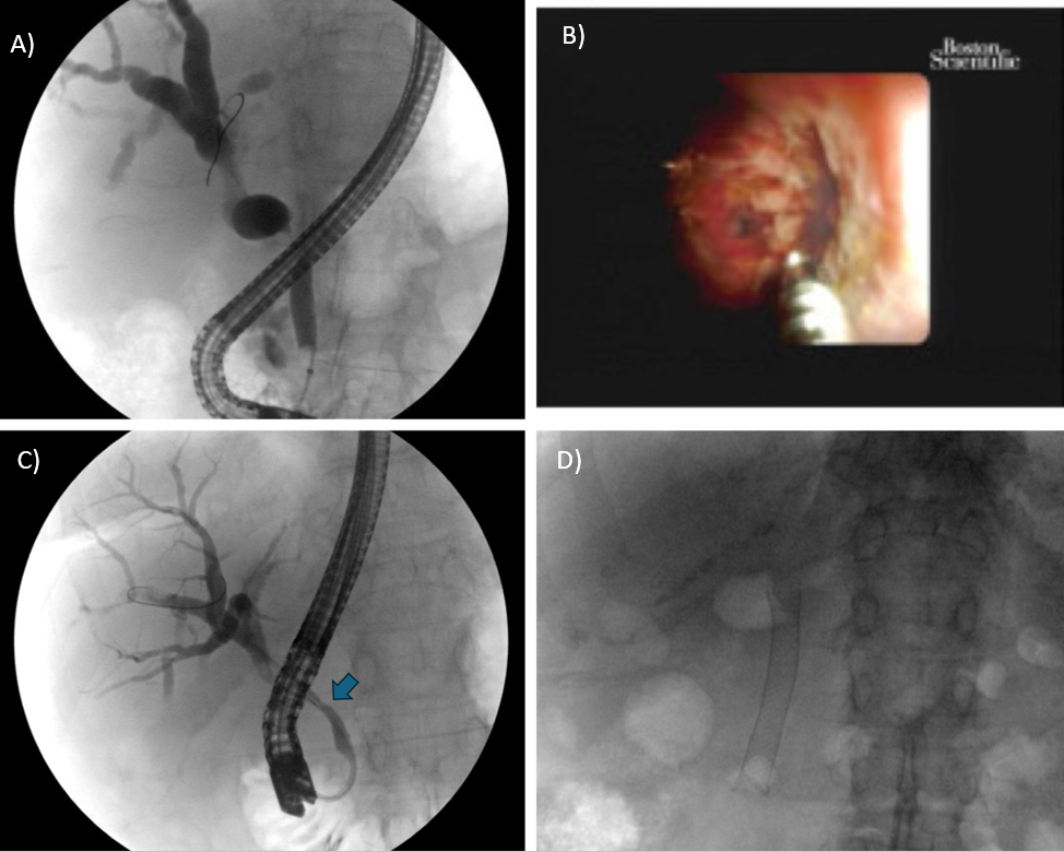
Methods: A 77-year-old female presented to the emergency room with obstructive jaundice, lab work showing AST 159 U/L, ALT 222 U/L, alkaline phosphatase 1,143 U/L, and total bilirubin 4 mg/dL. Abdominal ultrasound showed biliary ductal dilation, common bile duct (CBD) measuring 11.2 mm with diffuse intrahepatic ductal dilation. MRI demonstrated intrahepatic ductal dilation with middle CBD stenosis. Endoscopic ultrasound (EUS) revealed a middle CBD stricture which was sampled. EUS FNA revealed reactive ductal epithelial cells, low Ki67 proliferation, and no aberrant p53 expression, consistent with heterotopic pancreatic tissue. ERCP with spyglass cholangioscopy with spybite biopsy showed ectopic pancreatic tissue in the CBD. Patient was subsequently treated with intraductal radiofrequency ablation (RFA) and metal stent placement with complete resolution of her stricture and symptoms. Discussion: Embryologically, heterotopic pancreas occurs along the foregut due to the congruent development of primitive pancreatic tissue and foregut structures during week 4. Disruption of regional specific pancreatic transcription factors such as Hes1 and GATA4 alter pancreatic tissue organization and location specification. Specifically, misexpression of PTF1A, a transcription factor for pancreatic specification in the bile duct, has been linked with aberrant growth in the biliary tract. In this case, the ectopic pancreatic tissue created a biliary stricture, causing cholestatic liver injury which was diagnosed with EUS and ERCP and resolved with endoscopic RF treatment and metal stent placement. This case highlights the importance of understanding the most common sites of ectopic pancreatic tissue and the spectrum of its complications.
Figure: A) EUS showing biliary stricture within the CBD. B) Pathology of pancreatic acinar tissue biopsied.
Figure: A) ERCP with cholangiogram showing biliary structure. B) ERCP with spybite. C) ERCP with RFA. D) ERCP with metal stent placement.
Disclosures: Fady Gendy indicated no relevant financial relationships. Michael Makar indicated no relevant financial relationships. Veneeth Antony indicated no relevant financial relationships. Aws Alameri indicated no relevant financial relationships. Patrick Dorion indicated no relevant financial relationships. Bradley Confer indicated no relevant financial relationships. Harshit Khara indicated no relevant financial relationships.
Fady Gendy, DO1, Michael Makar, MD1, Veneeth Antony, DO1, Aws Alameri, MD1, Patrick Dorion, MD1, Bradley D. Confer, DO1, Harshit S. Khara, MD2. P2345 - Liver Blocked by Pancreas: An Unexpected Cause of a Biliary Stricture, ACG 2025 Annual Scientific Meeting Abstracts. Phoenix, AZ: American College of Gastroenterology.