Arkansas College of Osteopathic Medicine Fort Smith, AR
Muhammad Usman, MD, Muhammad G. Arnous, MD, Sanitha Pulapattassery, MD Arkansas College of Osteopathic Medicine, Fort Smith, AR Introduction: Haemobilia, or bleeding into the biliary tract, is a rare but a serious complication following liver biopsy. It develops due to abnormal communication between hepatic vasculature and biliary tract post liver biopsy. It often presents with gastrointestinal bleeding, jaundice, abdominal pain though all three are not always present simultaneously. We present a case of haemobilia and biliary tract obstruction after ultrasound guided liver biopsy in a patient of metastatic liver disease.
Case Description/
Methods: A 75-year-old female with a history of recently diagnosed colonic adenocarcinoma presents with acute right upper quadrant pain and upper GI bleed. She was recently evaluated for iron deficiency anemia with colonoscopy and found to have colonic adenocarcinoma with metastatic hepatic lesions on imaging. An ultrasound-guided liver biopsy was performed for metastatic evaluation about six days earlier. At presentation, she was found to be having significant elevation of liver function tests and bilirubin compared to normal levels before liver biopsy. CT scan of the abdomen revealed a lateral right hepatic lobe hemorrhage; distended gallbladder filled with echogenic debris and common bile duct dilated to 1.5 cm. Endoscopic retrograde cholangiopancreatography revealed multiple filling defects, followed by removal of multiple blood clots and placement of a biliary stent. A cholecystostomy drain was also placed for gallbladder echogenic debris, and right hepatic artery segmental branch embolization was performed for bleeding control. Following these interventions, abdominal pain gradually improved, and LFTs and serum bilirubin levels normalized. Patient was then discharged with outpatient follow-up. Her biliary stent and cholecystostomy drain were subsequently removed without any complications. Discussion: This case underscores the need to recognize haemobilia as a rare yet serious complication of liver biopsy. Although ultrasound-guided biopsies are considered relatively safe, they carry risks of vascular injury, particularly in patients with liver metastases or coagulopathies. Early recognition is challenging especially in people who do not present with classic triad of GI bleed, abdominal pain and jaundice. Therapeutic interventions including ERCP and arterial embolization in our patient lead to rapid improvement of patient's clinical condition. For patients undergoing liver biopsy, awareness of potential complications is crucial, especially in those with advanced malignancy or frailty.
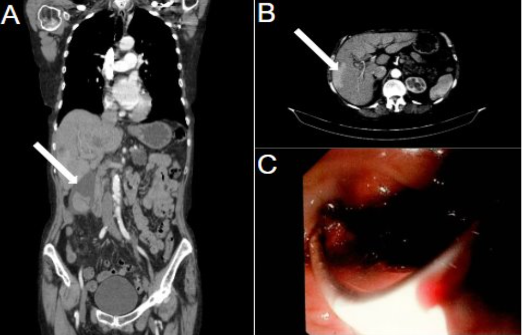
Figure: Figure A&B: CT scan of the abdomen with contrast showing distended gallbladder filled with echogenic debris and right hepatic lobe hemorrhage. Figure C: Endoscopic image showing blood clots at ampulla of water.
Disclosures: Muhammad Usman indicated no relevant financial relationships. Muhammad G. Arnous indicated no relevant financial relationships. Sanitha Pulapattassery indicated no relevant financial relationships.
Muhammad Usman, MD, Muhammad G. Arnous, MD, Sanitha Pulapattassery, MD. P2341 - Haemobilia and Biliary Tract Obstruction: A Rare Complication After Liver Biopsy, ACG 2025 Annual Scientific Meeting Abstracts. Phoenix, AZ: American College of Gastroenterology.

 photo")