
Monday Poster Session
Category: Colon
Navin Naik, DO
Franciscan Health Olympia Fields
Chicago, IL
Patient 1: A 61-year-old male with no significant medical history presented with abdominal pain, non-bloody dry heaving, and diarrhea. Imaging showed partial colonic obstruction, and he underwent an extended right hemicolectomy following failed conservative management. Pathology confirmed medullary carcinoma with MLH1 and PMS2 loss, but BRAF mutation testing was negative. He recovered after postoperative ileus and was discharged for outpatient oncology follow-up.
Patient 2: A 91-year-old female presented with abdominal pain and was found to have a cecal mass with abscess formation. Following surgical resection, pathology confirmed invasive medullary carcinoma with lymphatic invasion and rupture through the colon wall. Immunohistochemistry showed loss of MLH1 and PMS2 expression. The patient was started on Pembrolizumab based on subsequent PD-L1 testing.
Patient 3: An 86-year-old female with early satiety, nausea, and significant unintentional weight loss was found to have near-obstructing cecal cancer on colonoscopy. Biopsy confirmed medullary carcinoma. She underwent right hemicolectomy after nutritional stabilization and was referred for outpatient hematologic oncology follow-up.
Image 1 shows pathology from each patient.
Image 2 shows a negative stain from 2 patients.
Discussion: This case series underscores the diagnostic variability and therapeutic challenges associated with medullary carcinoma of the colon. While it predominantly affects elderly females, our series includes a younger male patient, highlighting potential atypical presentations. All patients showed loss of MLH1 and PMS2 expression, reinforcing the role of immunohistochemical profiling in diagnosis. Management often requires a multimodal approach, including surgical resection and immunotherapy. Early recognition and appropriate intervention are crucial given the unique biology and clinical behavior of this malignancy.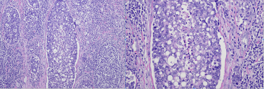
Figure: Image 1: Pathology slides showing poorly differentiated tumor cells consistent with medullary carcinoma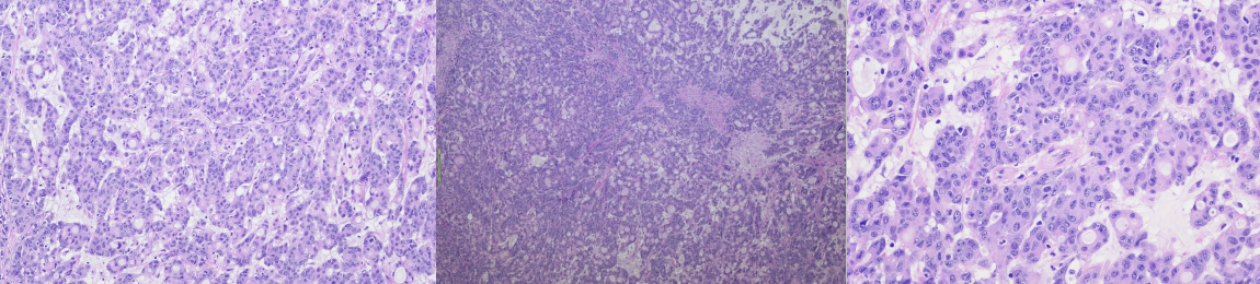
Figure: Image 2: MLH1 and PMS2 stains negative, consistent with medullary carcinoma
Disclosures:
Navin Naik indicated no relevant financial relationships.
Komlan Guedze indicated no relevant financial relationships.
Alexander Kadah indicated no relevant financial relationships.
Shadi Jibawi indicated no relevant financial relationships.
Navin Naik, DO1, Komlan Guedze, MD2, Alexander Kadah, MD1, Shadi Jibawi, MD3. P2476 - Medullary Carcinoma of the Colon: A Case Series Highlighting Diagnostic and Therapeutic Challenges, ACG 2025 Annual Scientific Meeting Abstracts. Phoenix, AZ: American College of Gastroenterology.