P2599 - Malignant Transformation of a Tailgut Duplication Cyst Into Squamous Cell Carcinoma: A Rare Cause of Perirectal Mass in a Patient With Ulcerative Colitis and Cirrhosis
Texas Tech University Health Sciences Center El Paso, TX
Omer Usman, MD1, Astin Worden, MD2, Thuy-Duyen Nguyen, MD2, Manreet Kaur, MBBS, MD2 1Texas Tech University Health Sciences Center, El Paso, TX; 2Mayo Clinic, Phoenix, AZ Introduction: Tailgut duplication cysts are rare congenital remnants of the embryonic hindgut, typically benign and incidentally discovered. Incidence is unknown, with only a few hundred cases reported globally. Malignancy rates of resected tail gut cysts reported in the literature are variable and range from 8% to 32%.Adenocarcinoma and neuroendocrine tumors are the most common malignancies, while transformation to squamous cell carcinoma (SCC) is rare and poorly characterized. We present a rare case of perirectal SCC arising from a tailgut cyst in a patient with extensive ulcerative colitis (UC) and well-compensated HCV-related cirrhosis.
Case Description/
Methods: A 66-year-old male with long-standing Ulcerative colitis in deep remission on mesalamine and compensated HCV cirrhosis with prior esophageal varices, presented with acute abdominal pain and diarrhea. Initially treated empirically for infectious enteritis, he returned with persistent pain. CT showed a 3.1 cm perirectal lesion; MRI revealed a 4.1 cm exophytic mass with central necrosis. Lower EUS identified a hypoechoicperirectal cystic mass with friable mucosa. FNA yielded scant fluid; cytology confirmed SCC. PET-CT demonstrated an FDG-avid mass without metastases. The lesion was suspected to arise from a tailgut cyst based on imaging and the absence of mucosal involvement. Due to rectal varices, surgery was deferred. The patient was managed with chemoradiotherapy with curative intent. Discussion: Tailgut cysts are rare developmental anomalies, more common in middle-aged women, with malignancy in < 10% of cases. SCC is a particularly rare subtype. This case is notable for the coexistence of UC and portal hypertension, complicating both diagnosis and treatment. Imaging and EUS suggested a duplication cyst, later confirmed malignant. No consensus exists for managing malignant tailgut cysts, underscoring the importance of multidisciplinary evaluation. Prompt recognition, staging, and oncologic management are critical for optimal outcomes.
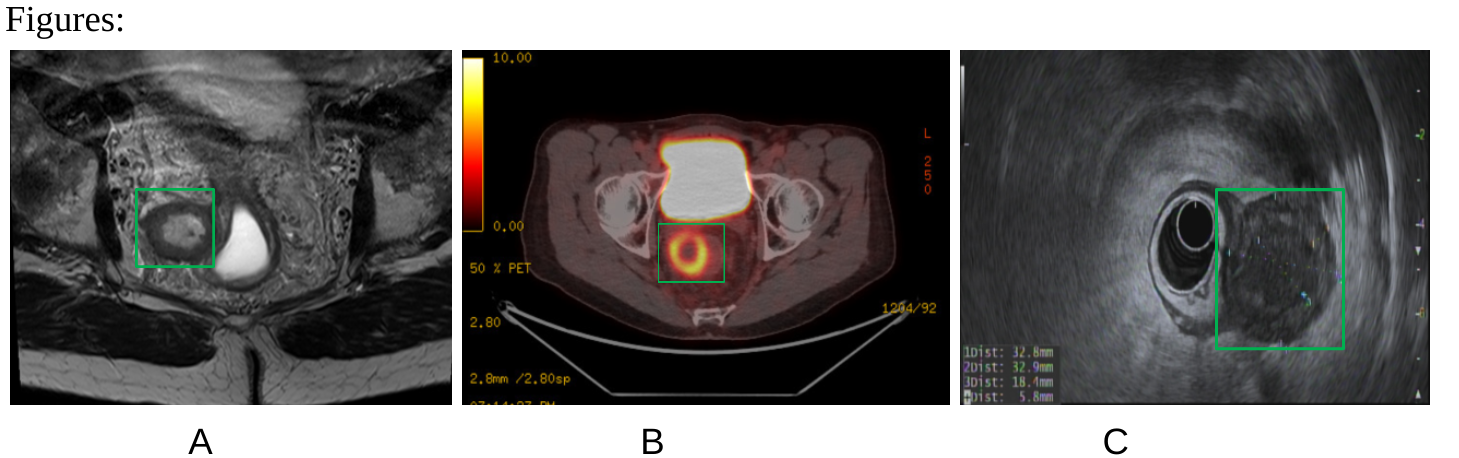
Figure: A: Non-contrast MRI pelvis showed a 4.1 cm exophytic lesion off the right mid-rectum with a thickened wall and probable central necrosis; differential includes necrotic GIST, atypical neoplasm, or chronic abscess. B: PET scan showed a centrally photopenic perirectal mass with a thickened wall, measuring 3.8 × 3.6 cm (4.1 × 3.2 cm on prior MRI). C: EUS showed a well-defined, hypoechoic perirectal cystic mass (33 × 33 × 5 mm). FNA (22G, 2 passes) yielded 0.2 mL fluid for cytology and cultures.
Disclosures: Omer Usman indicated no relevant financial relationships. Astin Worden indicated no relevant financial relationships. Thuy-Duyen Nguyen indicated no relevant financial relationships. Manreet Kaur indicated no relevant financial relationships.
Omer Usman, MD1, Astin Worden, MD2, Thuy-Duyen Nguyen, MD2, Manreet Kaur, MBBS, MD2. P2599 - Malignant Transformation of a Tailgut Duplication Cyst Into Squamous Cell Carcinoma: A Rare Cause of Perirectal Mass in a Patient With Ulcerative Colitis and Cirrhosis, ACG 2025 Annual Scientific Meeting Abstracts. Phoenix, AZ: American College of Gastroenterology.