Hayes Walker, MD1, Tyson Amundsen, MD1, Satheesh Nair, MD2 1UTHSC, Memphis, TN; 2Methodist LeBonheur Healthcare, Memphis, TN Introduction: Kaposi's sarcoma is a vascular tumor caused by infection with human herpes virus 8 and is considered an AIDS-defining condition. KS is classified into 4 types based upon the clinical circumstances. These types include classic, endemic, iatrogenic and AIDS-associated. The incidence of KS has decreased with the use of antiretroviral therapy. The most common extracutaneous site of KS is the GI tract. Gastrointestinal-KS is usually asymptomatic and therefore the majority of these cases remain undiagnosed. GI-KS is diagnosed by endoscopy with tissue biopsy. Combination ART is the standard of care for all AIDS-related KS. Initiation of systemic chemotherapy is considered when patients have advanced or rapidly progressive disease.
Case Description/
Methods: A 40-year-old African American male with PMHx of AIDS, hypertension, bipolar disorder, and depression presented to the ED for several days of RUQ abdominal pain, shortness of breath, chest pain, alternating bloody diarrhea and constipation, as well as fever. He was recently seen at an urgent care and given amoxicillin for sinusitis. He reported compliance with Biktarvy, however, he was not taking recently prescribed Bactrim. Most recent CD4 count was 36. He had recently received a dose of Bicillin for a positive RPR. Imaging was significant for right lower lobe pneumonia and low-grade enterocolitis. GI pathogen panel was positive for Campylobacter, Shigella/Enteroinvasive E. coli, and Giardia. EGD revealed a bleeding, fungating 2 cm mass with malignant appearance in the body of the stomach. A villous, nonbleeding 1 cm mass with a benign appearance was seen in the fundus. A few localized erosions were also seen in the second part of the duodenum. Biopsied sections of the malignant-appearing mass showed ulcerated gastric mucosa with underlying vascular proliferation in the lamina propria with areas of spindle cells and prominent red blood cell extravasation. Immunohistochemistry was positive for HHV-8 and CD34 and notably negative for CK Oscar, CMV and HSV. The patient was discharged home on ART and appropriate antibiotics. Discussion: Endoscopy with biopsy allows for immunohistochemical testing to confirm the diagnosis of GI-KS. Treatment includes antiretroviral therapy +/- systemic chemotherapy, such as liposomal doxorubicin, in the setting of advanced or progressive disease. Future studies are needed to investigate the utility of performing screening endoscopies in select patients for early detection and treatment of GI-KS.
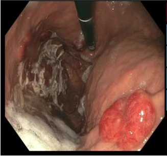
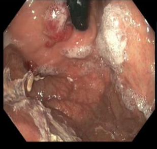
Figure: Image A: Bleeding, fungating 2 cm mass with malignant appearance in the body of the stomach.
Figure: Image B: Villous, nonbleeding 1 cm mass with a benign appearance seen in the fundus.
Disclosures: Hayes Walker indicated no relevant financial relationships. Tyson Amundsen indicated no relevant financial relationships. Satheesh Nair indicated no relevant financial relationships.
Hayes Walker, MD1, Tyson Amundsen, MD1, Satheesh Nair, MD2. P3485 - A Case of Gastrointestinal Kaposi's Sarcoma, ACG 2025 Annual Scientific Meeting Abstracts. Phoenix, AZ: American College of Gastroenterology.