Baylor Scott & White Medical Center Round Rock, TX
Neel Shah, MD, Rahul Thakkar, MD, Nathan Mielke, DO Baylor Scott & White Medical Center, Round Rock, TX Introduction: Endometriosis is a chronic inflammatory disorder characterized by the presence of functional endometrial tissue outside the uterine cavity. Hepatic endometriosis is exceedingly rare and often mimics hepatic neoplasms on imaging. We present a unique case of intrahepatic endometriosis, incidentally discovered after a motor vehicle collision.
Case Description/
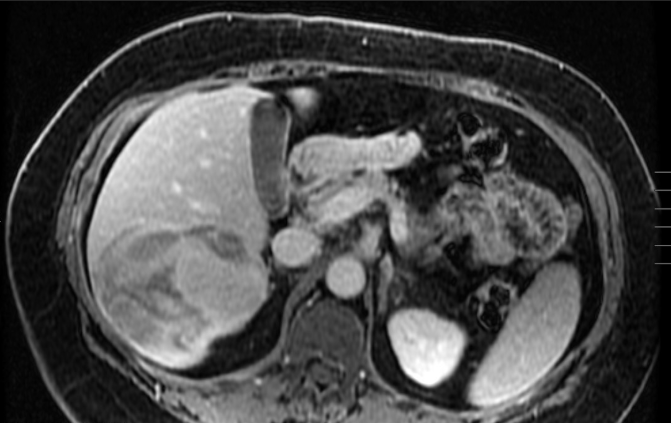
Methods: A 54-year-old woman with a history of laparoscopic resection of an ovarian endometrioma in 2017, class I obesity, and prior bariatric surgery was evaluated for a hepatic lesion incidentally discovered after a motor vehicle collision in 2022. Initial CT imaging revealed a heterogeneous solid and cystic mass in the right hepatic lobe measuring 8.2 × 4.5 cm, along with a peritoneal nodule in the left anterior abdominal wall measuring up to 1.2 cm—both initially presumed to be traumatic hematomas. A follow-up MRI noted a 4 cm right hepatic lesion consistent with an intraparenchymal hematoma. After the patient obtained insurance coverage, a repeat MRI in March 2025 showed a complex right hepatic lesion measuring 8.9 × 8.0 cm with internal blood products and peripheral enhancement (Figure 1), as well as an enlarging anterior peritoneal nodule measuring 2.2 × 1.2 cm. Due to concern for malignancy, the patient underwent an ultrasound-guided liver biopsy. Histopathological analysis demonstrated hepatic parenchyma with smooth muscle hyperplasia, hemorrhage, and focal epithelial proliferation. Immunohistochemical staining was positive for PAX8, cytokeratin 7 (CK7), and estrogen receptor (ER), consistent with intrahepatic endometriosis. Discussion: Intrahepatic endometriosis is an exceptionally rare condition, with fewer than 30 cases reported worldwide. Its imaging appearance can mimic primary or metastatic hepatic tumors, making diagnosis challenging. This case highlights the importance of maintaining a broad differential in patients with atypical hepatic lesions, particularly those with a history of endometriosis, and underscores the critical role of histopathology and immunohistochemistry in establishing a definitive diagnosis.
Figure: Figure 1: MRI of the liver showing a cystic heterogenous right lobe liver mass
Disclosures: Neel Shah indicated no relevant financial relationships. Rahul Thakkar indicated no relevant financial relationships. Nathan Mielke indicated no relevant financial relationships.
Neel Shah, MD, Rahul Thakkar, MD, Nathan Mielke, DO. P3962 - A Rare Hepatic Mass Revealed as Intrahepatic Endometriosis, ACG 2025 Annual Scientific Meeting Abstracts. Phoenix, AZ: American College of Gastroenterology.

.jpg "Neel Shah, MD (he/him/his) photo")