Alexander Dile, MD, Patrick J. Carey, MD, Jonathan S. Moulton, MD, Milton Smith, MD University of Cincinnati, Cincinnati, OH Introduction: Polycystic kidney disease (PKD) often involves other organ systems including the liver. While most cases have little impact on the liver's synthetic function, this condition can significantly alter the hepatic architecture thus obscuring other conditions. We present a case of metastatic pancreatic cancer with liver metastasis which was initially concealed behind a background of benign cysts.
Case Description/
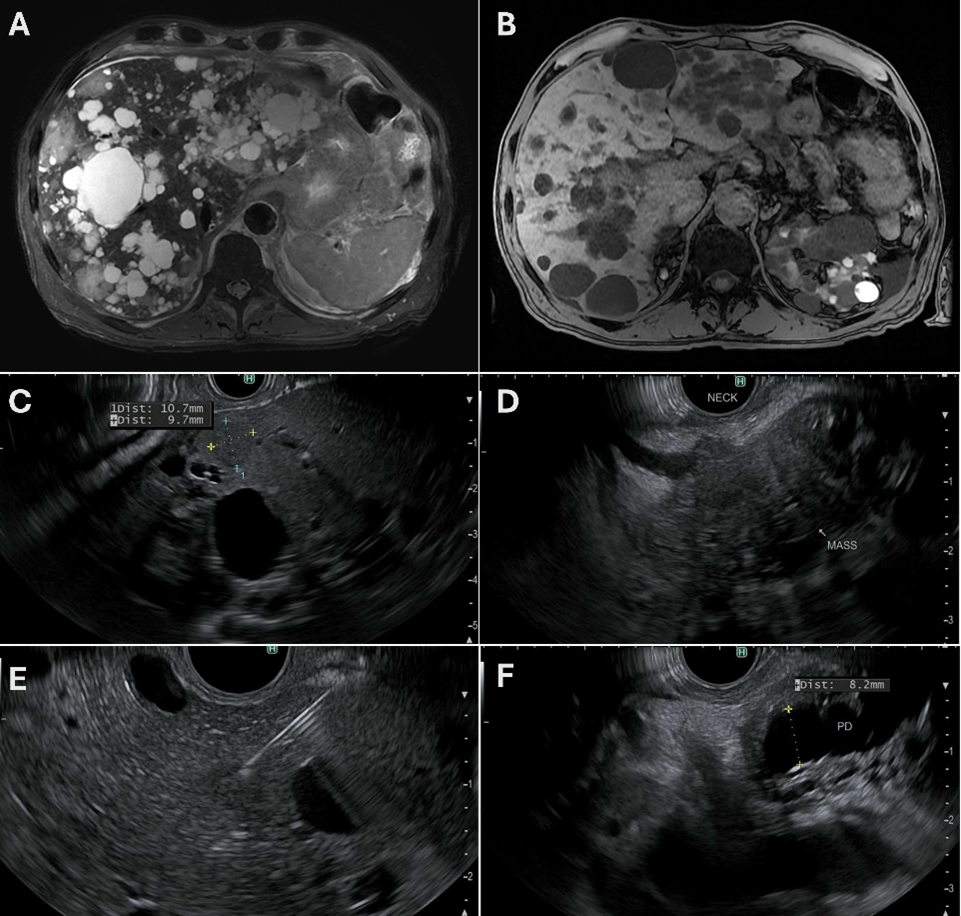
Methods: A 66-year-old male with a kidney transplant three years prior due to PKD and diabetes presents to the emergency department with recurrent fevers. He was recently hospitalized for klebsiella bacteremia of unknown origin and discharged on appropriate antibiotics but represented several days later with persistent fevers and abdominal pain. His labs were notable for a WBC 12.1K, ALP 198g/dL, and CRP 123mg/L. CT abdomen and pelvis showed extensive polycystic disease involving the liver and pancreas. An infectious workup including bacterial, fungal and viral cultures, serologies and CSF analyses were all negative. PET CT imaging showed multiple foci of increased FDG activity throughout the liver and pancreatic body. Subsequent MRI identified innumerable non-cystic hepatic lesions and an ill-defined pancreatic body mass. Endoscopic ultrasound showed an ill-defined hypoechoic mass in the pancreatic neck with upstream pancreatic ductal dilation and a solid mass in the liver, both of which were biopsied (Figure 1). Pathology returned positive for adenocarcinoma. Discussion: This case highlights the challenges in diagnosing underlying malignancies in patients with complex anatomic abnormalities, such as those with PKD, and the importance of inter-specialty collaboration along with utilization of advanced endoscopic techniques to confirm diagnosis.
Figure: A) Axial T2 non-contrast MRI showing many mildly hyperdense focal hepatic lesions surrounded by innumerable cystic lesions. B) Axial Dual Echo MRI showing widespread hepatic cysts. C) EUS of a right liver lobe mass measuring approximately 10.7 mm x 9.7mm. D) EUS of an ill-defined pancreatic neck mass. E) EUS showing fine needle biopsy of the right liver lobe lesion. F) EUS showing pancreatic ductal dilation measuring up to 8.2mm.
Disclosures: Alexander Dile indicated no relevant financial relationships. Patrick Carey indicated no relevant financial relationships. Jonathan Moulton indicated no relevant financial relationships. Milton Smith indicated no relevant financial relationships.
Alexander Dile, MD, Patrick J. Carey, MD, Jonathan S. Moulton, MD, Milton Smith, MD. P3946 - Hidden in Plain Sight: A Polycystic Liver Concealing Metastatic Pancreatic Cancer, ACG 2025 Annual Scientific Meeting Abstracts. Phoenix, AZ: American College of Gastroenterology.

 photo")