Venkat Arutla, MD1, Ese Uwagbale, MD2, Karin Dunnigan, MD1 1Rochester General Hospital, Rochester, NY; 2Rochester General Hospital, Webster, NY Introduction: Unintentional weight loss is a daunting symptom that often requires extensive investigations to rule out possible underlying cancer. Gastrointestinal (GI) amyloidosis occurs when insoluble protein deposits along the GI tract, causing altered function. Although well-known throughout the literature, GI amyloidosis may often be overlooked in the differential diagnosis for weight loss. Here, we present a case of GI amyloidosis presenting with weight loss.
Case Description/
Methods: A 69-year-old female with a past medical history of morbid obesity status post Roux-en-Y gastric bypass and osteoporosis presented to her primary care provider with 40 pounds of weight loss, nausea, and vomiting of 2 months duration.
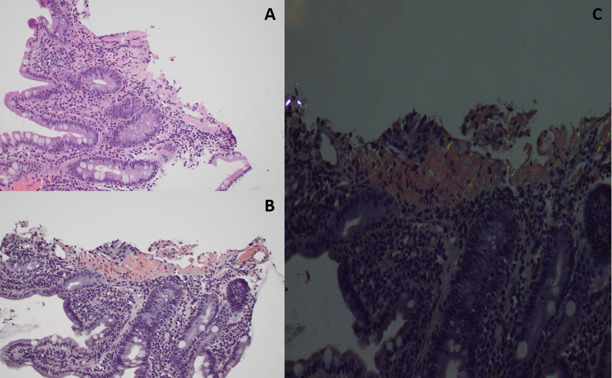
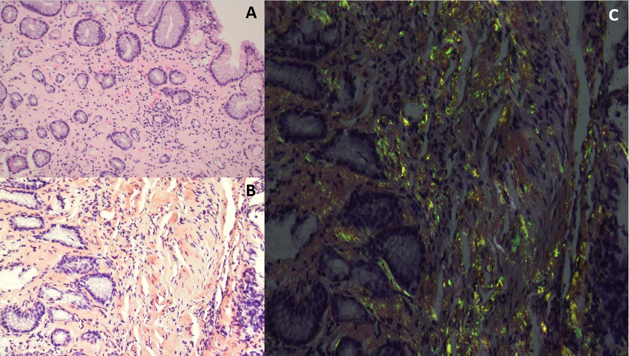
Computed tomography (CT) of the abdomen and pelvis showed a moderate-sized para-esophageal hernia with a partially herniated gastric pouch. Esophagogastroduodenoscopy (EGD) revealed a 5 mm, acute-appearing, punched-out ulceration in the jejunum, seen 9 cm from the gastrojejunal anastomosis. Biopsies of the ulcer and the stomach revealed small intestinal and gastric mucosa with a positive Congo red stain, indicating amyloid deposition (Figure 1 and 2). Amyloid protein identification by mass spectroscopy showed Amyloidosis AL (kappa)-type.
There was also a concern for systemic amyloidosis with renal and cardiac involvement as the patient also had acute kidney injury with a creatinine of 1.4, and an echocardiogram showed a severely dilated left atrium.
The patient was referred to hematology, and she was started on systemic therapy with daratumumab, cyclophosphamide, bortezomib, and dexamethasone. Discussion: The differential diagnosis for weight loss is broad; infiltrating systemic diseases, such as amyloidosis, should be considered as a possible differential diagnosis.
Pathology slides courtesy of Dr. Fadi Hatem (pathology department Rochester General Hospital).
Figure: Figure 1: Jejunum biopsy showing routine hematoxylin & eosin stain (A), positive Congo red stain (B) and polarized Congo red stain (C).
Figure: Figure 2: Stomach biopsy showing routine hematoxylin & eosin stain (A), positive Congo red stain (B) and polarized Congo red stain (C).
Disclosures: Venkat Arutla indicated no relevant financial relationships. Ese Uwagbale indicated no relevant financial relationships. Karin Dunnigan indicated no relevant financial relationships.
Venkat Arutla, MD1, Ese Uwagbale, MD2, Karin Dunnigan, MD1. P4088 - A Case of Gastrointestinal Amyloidosis Presenting With Weight Loss, ACG 2025 Annual Scientific Meeting Abstracts. Phoenix, AZ: American College of Gastroenterology.