Louisiana State University School of Medicine New Orleans, LA
Award: ACG Presidential Poster Award
Farhan Mohiuddin, MD1, Katherine Westbrook Cates, DO1, Catherine Hudson, MD, MPH2, Jason Stibbe, MD3 1Louisiana State University School of Medicine, New Orleans, LA; 2Louisiana State University Health, New Orleans, LA; 3LSU Health New Orleans School of Medicine, New Orleans, LA Introduction: Lobular capillary hemangioma (LCH), also known as pyogenic granuloma, is a benign vascular tumor often arising in the skin or mucous membranes. Epidemiologic data is conflicting, with some studies showing a male predominance in the 20s, others noting female predominance in the 30s–40s, and others reporting higher incidence in pediatric patients. The etiology is unclear, with theories including trauma, infection, vascular malformations, medications, or estrogen-associated inflammation. While LCH can occur at any cutaneous or mucosal site, cutaneous lesions are more common. Mucosal cases typically affect the lips and gingiva. Gastric involvement is rare, especially in the gastric body. Immunosuppressed patients, such as those post-transplant, have increased susceptibility. We present a rare case of gastric body LCH presenting with hematemesis.
Case Description/
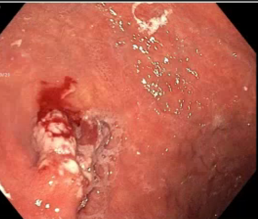
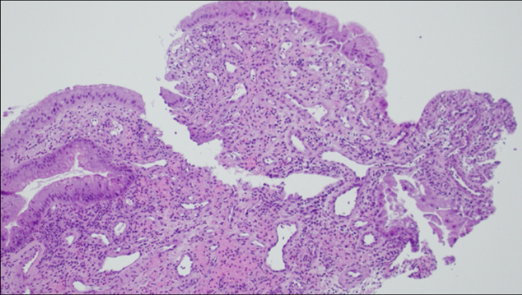
Methods: A 74-year-old male with atrial fibrillation on Eliquis, hypertension, hyperlipidemia, and osteoarthritis presented with large-volume hematemesis and transient unresponsiveness while fishing. He was hemodynamically stable on arrival. He had recently been diagnosed with anemia and was awaiting outpatient endoscopic evaluation. Admission labs showed hemoglobin of 9.9, MCV 78.9, RDW 19.6, and BUN 26. EGD revealed a 2x3 cm indurated lesion with surrounding clots in the gastric fundus. Biopsies were taken and Hemospray was applied. Post-EGD, he remained stable and was discharged with anticoagulation discontinued after shared decision-making. Pathology revealed focal vascular proliferation consistent with lobular capillary hemangioma. He is clinically stable on outpatient follow-up and plans surgical resection to prevent recurrent bleeding and improve anemia. Discussion: Gastric LCH is exceedingly rare. Few cases describe LCH in the gastric antrum or duodenal bulb presenting with anemia. Treatments have included hot snare removal, surgery, or radiofrequency ablation. Proposed mechanisms include angiogenic dysregulation and associations with medications like immunosuppressants, contraceptives, chemotherapy, retinoids, or antiretrovirals—none of which were present in this patient. To our knowledge, this is the first reported case of gastric body LCH presenting with hematemesis. Surgical resection may be warranted in cases of bleeding or refractory anemia. More data is needed to guide diagnosis and management.
Figure: Endoscopic image of lobar capillary hemangioma located in the gastric fundus.
Figure: Histopathological biopsy specimen of the gastric fundus mass consistent with lobar capillary hemangioma.
Disclosures: Farhan Mohiuddin indicated no relevant financial relationships. Katherine Westbrook Cates indicated no relevant financial relationships. Catherine Hudson indicated no relevant financial relationships. Jason Stibbe indicated no relevant financial relationships.
Farhan Mohiuddin, MD1, Katherine Westbrook Cates, DO1, Catherine Hudson, MD, MPH2, Jason Stibbe, MD3. P4224 - A Rare Case of Hematemesis: Lobular Capillary Hemangioma in the Gastric Body, ACG 2025 Annual Scientific Meeting Abstracts. Phoenix, AZ: American College of Gastroenterology.


.jpg "Farhan Mohiuddin, MD photo")