University of Texas Rio Grande Valley Edinburg, TX
Kashif Ali, MD1, Jaya Vasudevan, MD2, Shuhaib Ali, DO2, Prabhleen Chahal, MD, FACG2 1University of Texas Rio Grande Valley, Edinburg, TX; 2University of Texas Health San Antonio, San Antonio, TX Introduction: Splenosis is the ectopic presence of splenic tissue in abnormal locations, typically following splenic trauma or splenectomy. It is usually asymptomatic and discovered incidentally on imaging. While most often found in intraperitoneal or intrathoracic locations, splenosis occurring in the deep retroperitoneal pelvic space is extremely rare and can mimic neoplastic or vascular lesions. Diagnosis can be challenging with cross-sectional imaging alone, and confirmation often requires histologic evaluation. We present a case of splenosis in the retroperitoneal pelvic space which highlights the value of endoscopic ultrasound (EUS) in the evaluation of an ectopic splenic mass in a patient with prior history of splenectomy.
Case Description/
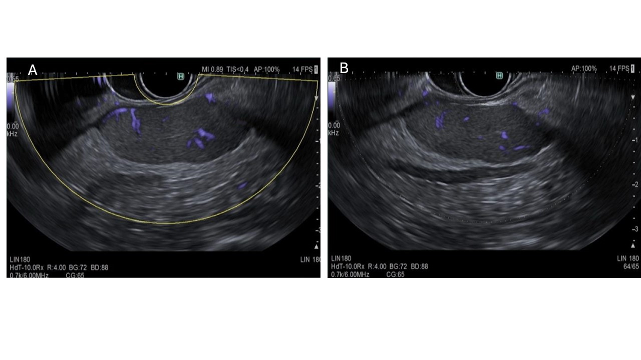
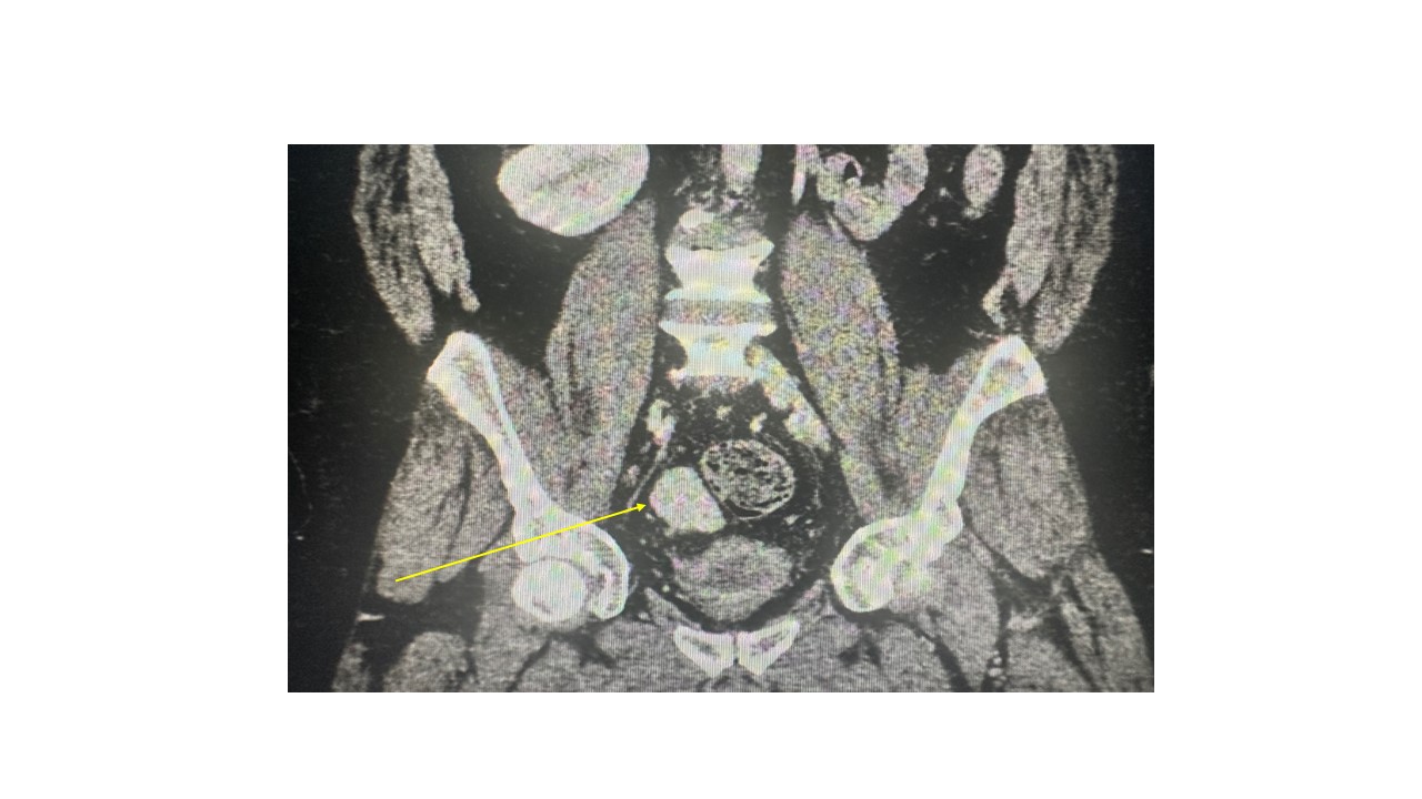
Methods: A 43-year-old male with a history of hypertension and remote motor vehicle accident resulting in left nephrectomy and splenectomy was initially referred to General Surgery by Oncology for evaluation of an asymptomatic right suprarenal retroperitoneal mass incidentally found on routine CT imaging. Imaging revealed a 10 cm retroperitoneal mass and a 7.2 × 2.8 cm oblong, lobulated hyperattenuating structure in the right hemipelvis, initially suspected to be a venous varix. The lack of definitive vascular communication led General Surgery to consult GI for further evaluation via EUS. EUS revealed a well-defined, isoechoic mass in the perirectal retroperitoneal space measuring 72 mm × 15 mm. Doppler imaging showed no significant vascularity. Based on echotexture and appearance, splenosis was suspected. Fine-needle aspiration was performed, and histopathology confirmed the diagnosis of splenic tissue. Given the patient’s lack of symptoms, the mass was managed conservatively. He later underwent unrelated surgical removal of a myelolipoma of the adrenal gland. Discussion: As underscored by this case, in patients with history of splenic trauma or splenectomy, splenosis may be considered in the differential diagnosis retroperitoneal masses. While splenosis is most often found in common intraperitoneal sites, its presence in the deep pelvic retroperitoneum is rare and easily misidentified. Cross-sectional imaging alone is often inconclusive, particularly in uncommon locations. In this case, EUS provided high-resolution visualization and real-time Doppler assessment, allowing targeted biopsy and definitive diagnosis without surgical intervention. The case emphasizes how EUS can be a powerful, minimally invasive tool in evaluating challenging retroperitoneal masses.
Figure: EUS showing an ectopic spleen in the rectum.
Figure: A CT scan showing an ectopic spleen in the rectum.
Disclosures: Kashif Ali indicated no relevant financial relationships. Jaya Vasudevan indicated no relevant financial relationships. Shuhaib Ali indicated no relevant financial relationships. Prabhleen Chahal indicated no relevant financial relationships.
Kashif Ali, MD1, Jaya Vasudevan, MD2, Shuhaib Ali, DO2, Prabhleen Chahal, MD, FACG2. P4272 - Rectal Mass or Something Else? A Rare Case of Ectopic Spleen in the Rectum and Diagnosed by Endoscopic Ultrasound, ACG 2025 Annual Scientific Meeting Abstracts. Phoenix, AZ: American College of Gastroenterology.