Ridhima Kaul, MD1, Arjun Chatterjee, MD1, Tyler Stevens, MD1, Hassan Siddiki, MD1, Amit Bhatt, MD1, Kyungran Justina Cho, MD, PhD2 1Cleveland Clinic Foundation, Cleveland, OH; 2Cleveland Clinic Foundation, Pepper Pike, OH Introduction: Hydro-Computed tomography (CT), a technique commonly used in Korea for staging early gastric cancer, may offer superior visualization of gastric wall lesions compared to traditional CT. Its application, as demonstrated in our case, highlights its potential utility in the United States.
Case Description/
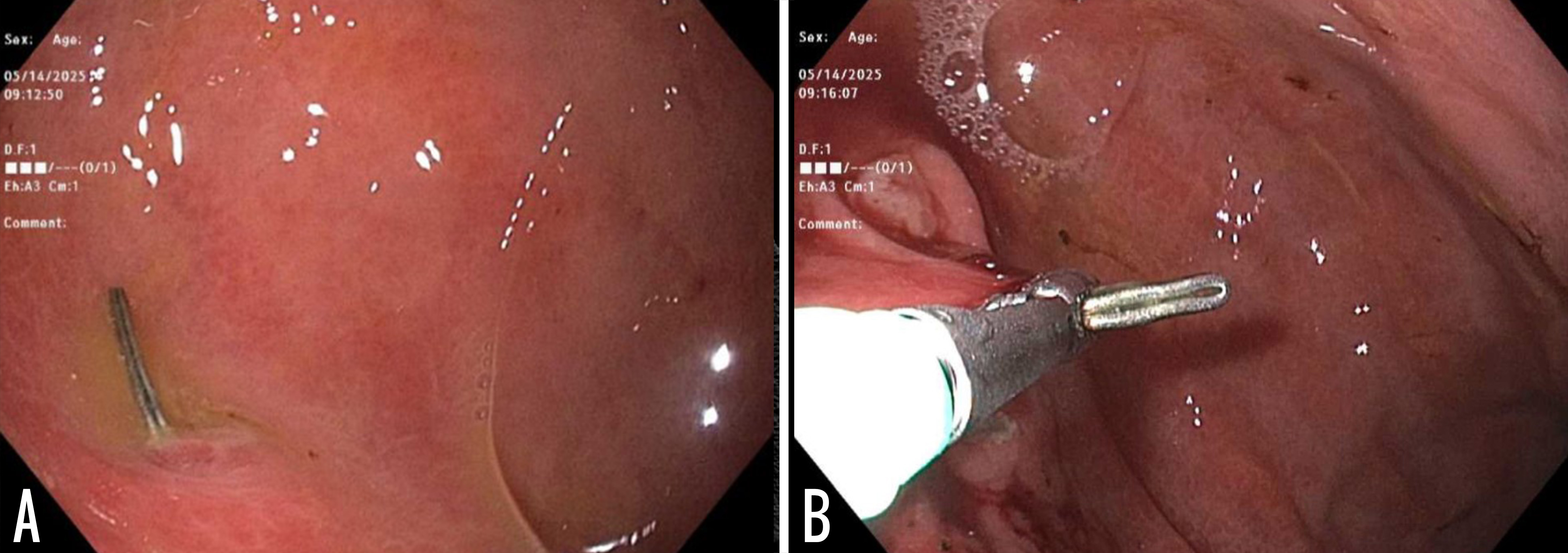
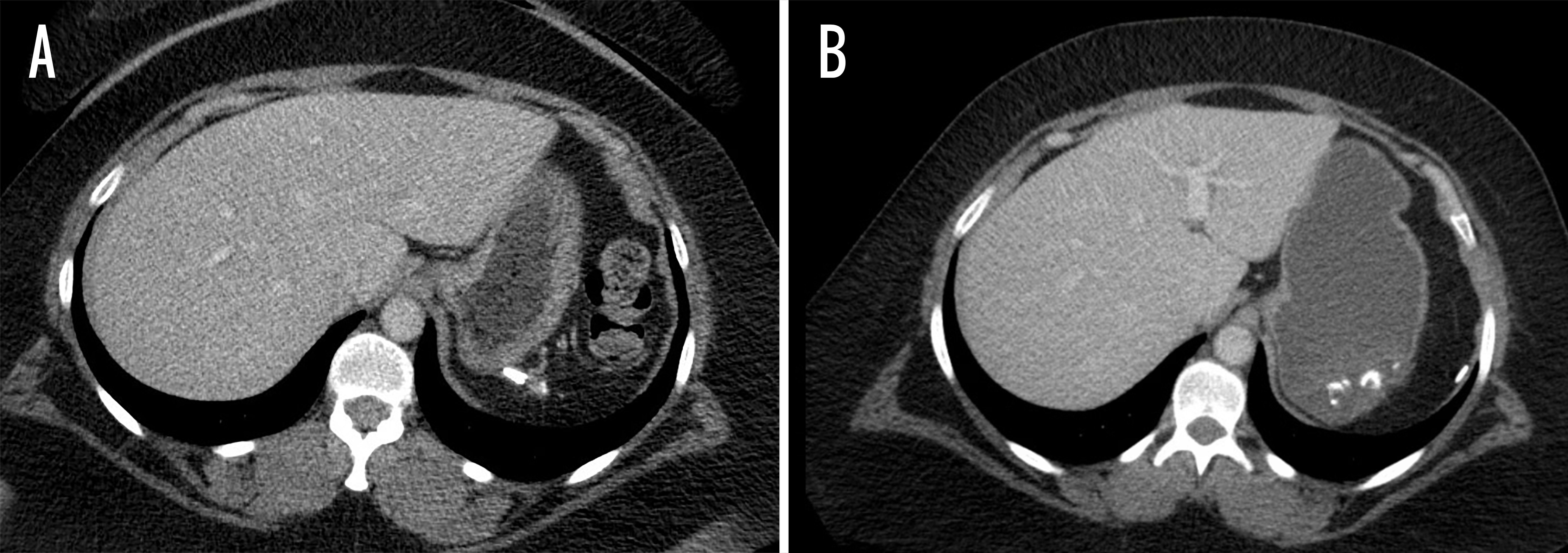
Methods: A 42-year-old female presenting with a past medical history of systemic lupus erythematosus complicated by immune thrombocytopenic purpura, status post splenectomy in 2001, presented to our hospital with a chief complaint of a three-day history of constant chest pain radiating to her left side with intermittent sharp shooting chest pains. Cardiac workup was negative for acute coronary syndrome. CT abdomen and pelvis showed two surgical clips related to her prior splenectomy located in the posterior wall of the stomach with potential erosion into the gastric wall, with associated focal thickening in the area of prior splenectomy (Figure 1a). It was unclear from this imaging whether the surgical clip was extrinsic to the posterior gastric wall or had eroded into the posterior wall of the gastric fundus. Gastroenterology was consulted for further recommendations. We recommended a helical hydro CT of the abdomen - a technique that uses water for distention of the stomach for better visualization of the area. Repeat imaging using the hydro-CT protocol showed a surgical clip within the lumen of the gastric fundus and an additional surgical clip at the ileocecal junction (Figure 1b). She underwent upper endoscopy with successful removal of the surgical clip within the stomach lumen (Figure 2a, b). This case presents a rare complication post-splenectomy: the migration of a surgical clip into the posterior gastric wall. Discussion: While a traditional CT of the abdomen with contrast could not determine of the if the clip was within or external to the gastric wall. The Hydro-CT was able to demonstrate that the clip was within the lumen of the stomach, allowing for appropriate endoscopic planning. Our case highlights the utility of the hydro-CT for better visualization of the gastric wall in the United States.
Figure: Figure 1. (A) Computed tomography (CT) of the abdomen showing surgical clips located along the posterior wall of the stomach, with question of possible erosion of the staples into the gastric wall. (B) Hydro-CT scan of the abdomen revealing a single surgical clip within the lumen of the gastric fundus.
Figure: Figure 2 (A.B) Upper endoscopy showing a clip in the fundus of the stomach, which was removed successfully.
Disclosures: Ridhima Kaul indicated no relevant financial relationships. Arjun Chatterjee indicated no relevant financial relationships. Tyler Stevens indicated no relevant financial relationships. Hassan Siddiki: Boston Scientific – Consultant. Conmed – Consultant. Amit Bhatt indicated no relevant financial relationships. Kyungran Justina Cho indicated no relevant financial relationships.
Ridhima Kaul, MD1, Arjun Chatterjee, MD1, Tyler Stevens, MD1, Hassan Siddiki, MD1, Amit Bhatt, MD1, Kyungran Justina Cho, MD, PhD2. P4264 - Utility of Hydro-CT in Diagnosing Surgical Clip Migration Into the Gastric Wall Post-Splenectomy, ACG 2025 Annual Scientific Meeting Abstracts. Phoenix, AZ: American College of Gastroenterology.

 photo")