Nick Adimi, MD, MS, BS1, Thomas Mientus, MD2, Meghana Doniparthi, MD1 1Advocate Lutheran General, Park Ridge, IL; 2GI Alliance, Midwest, Waukegan, IL Introduction: Gastric xanthomas are benign lesions that can be seen incidentally on endoscopy, characterized by a white-yellow plaque appearance. The reported incidence is 0.23-7% of people and noted rising incidence with advancing age. Recognition of these lesions is crucial, as there is a known association with gastric cancer. In this case, we discuss the implications of gastric xanthoma in a patient with a family history of gastric cancer, the importance of relevant conditions, and our recommendations for surveillance EGD.
Case Description/
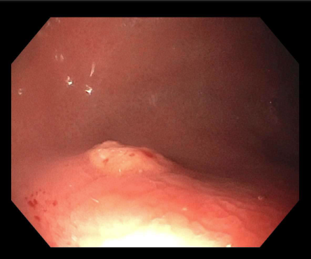
Methods: Our patient is a 38-year-old male with a family history of BRCA2 mutation in his father and gastric cancer in both his paternal grandfather and father. He was found to also have positive BRCA2 mutation and presented for screening endoscopy due to his history. An EGD 9 years prior for epigastric discomfort was reportedly normal. EGD done at this recent visit revealed antrum mucosal erythema and a 5 mm yellow polypoid lesion in the fundus which was removed with cold snare (Fig 1). Random biopsies were taken from the antrum and body separately, which were negative for intestinal metaplasia or H. pylori. The polyp pathology showed hyperplastic mucosa with characteristic foamy macrophages on CD68 immunostain consistent with xanthoma (Fig 2). Discussion: Gastric xanthomas are lipid-laden plaques or nodules that may present endoscopically as single or multifocal lesions with average size between 2-10 mm. Often these are asymptomatic lesions, but there are reports of early satiety, weight loss, or hematemesis if they grow larger in size. These generally arise in abnormal mucosa, such as atrophic gastritis, gastric intestinal metaplasia, and H. pylori infection. The prevalence of gastric xanthomas with these premalignant conditions may explain some of the reported association with development of gastric cancer; however, xanthomas are also an independent predictor of rapid tumor growth rate and recurrence after endoscopic resection. There is no formal endoscopic surveillance guideline for patients with gastric xanthoma. Our patient had multiple risk factors for gastric cancer: BRCA2, family history in two family members without hereditary diffuse gastric cancer criteria or gene mutations, and gastric xanthoma itself. In this case, we discussed surveillance EGD every 1-3 years.
Figure: Figure 1) 5 mm yellow polypoid lesion seen at the gastric fundus on endoscopy.
Figure: Figure 2) A: CD68 immunostain showing increased macrophage density as seen in gastric xanthoma. B: Pathology slide showing foamy macrophages seen in gastric xanthoma.
Disclosures: Nick Adimi indicated no relevant financial relationships. Thomas Mientus indicated no relevant financial relationships. Meghana Doniparthi indicated no relevant financial relationships.
Nick Adimi, MD, MS, BS1, Thomas Mientus, MD2, Meghana Doniparthi, MD1. P4241 - Case of Gastric Xanthoma: Examining Its Association With Gastric Cancer and Determining Surveillance EGD Timeframe, ACG 2025 Annual Scientific Meeting Abstracts. Phoenix, AZ: American College of Gastroenterology.