Ross M. Dies, MD, MS1, Farhan Mohiuddin, MD2, Brooke McVaney, DO3, Jessica Bordes, MD1 1Louisiana State University Health, New Orleans, LA; 2Louisiana State University School of Medicine, New Orleans, LA; 3LSU Health New Orleans, New Orleans, LA Introduction: Gastrointestinal stromal tumors (GISTs) are mesenchymal tumors of the gastrointestinal (GI) tract, most commonly occurring in the stomach or small intestine. Clinical manifestations may be non-specific, varying from asymptomatic to abdominal pain, palpable mass, GI bleeding, occlusion, and even perforation. GISTs often have solid tumor components on imaging but may also have cystic features. Cystic components normally occur with higher-grade malignancy, and these radiographic findings may resemble other pathologies, prompting alternative workup and treatment.
Case Description/
Methods: A 53-year-old woman with a history of gastroesophageal reflux disease and prior H. pylori infection presented to our center for outpatient endoscopy to monitor for resolution of a recently identified gastric ulcer. Patient had recently been admitted elsewhere for several weeks of abdominal pain and nausea; imaging revealed a gastric perforation and adjacent air-fluid collection. Antibiotics were administered, and a drain was placed at that time and removed weeks later. Endoscopy present-day revealed a cratered gastric ulcer oozing blood and draining purulent fluid, and patient was admitted with concern for gastric abscess. Repeat abdominal imaging showed an ulcerative exophytic fundal mass with mixed soft tissue and fluid attenuation, concerning for GIST, with scattered peritoneal nodules concerning for drop metastases. Endoscopic ultrasound (EUS) with fine needle aspiration was performed, and pathology results confirmed our suspicions for GIST. Patient was discharged on appropriate pharmacological therapy with specialist follow-up. Discussion: GISTs have varying radiographic features, with cystic degeneration and focal hemorrhage being present in larger, more advanced tumors. Larger GIST lesions with cystic spaces may communicate with the gastric lumen and demonstrate air-fluid levels on abdominal imaging. While this may resemble infectious collections, bacterial gastric wall abscess is an extremely rare phenomenon which is usually secondary to spread of another primary systemic infection or local invasion of the gastric mucosa. Therefore, visualization of fluid collections adjacent to the stomach wall should prompt evaluation for this, but also other pathologies.
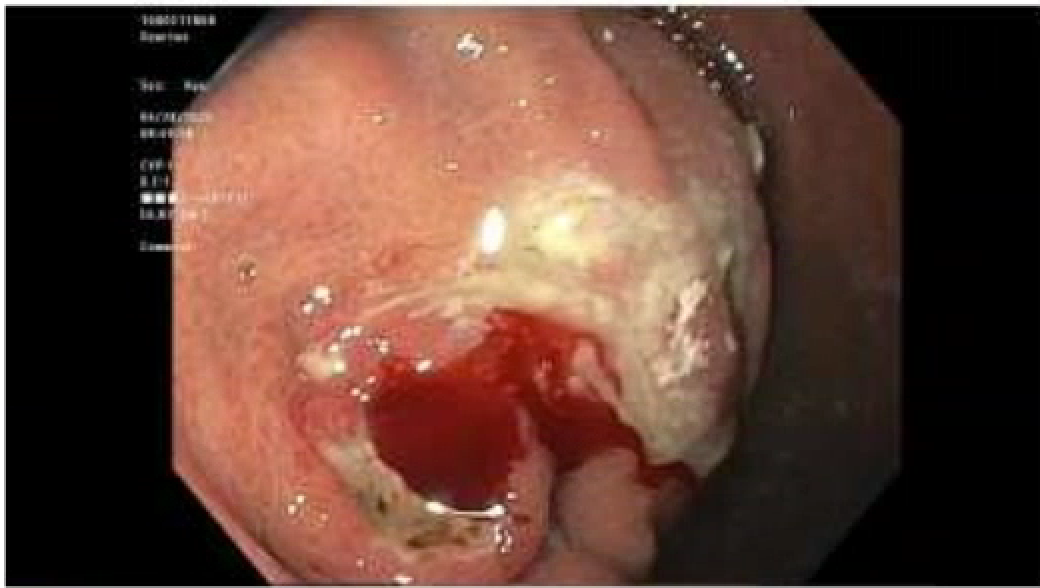
Figure: Visualization of bleeding ulcer in the gastric fundus with accompanying purulent drainage
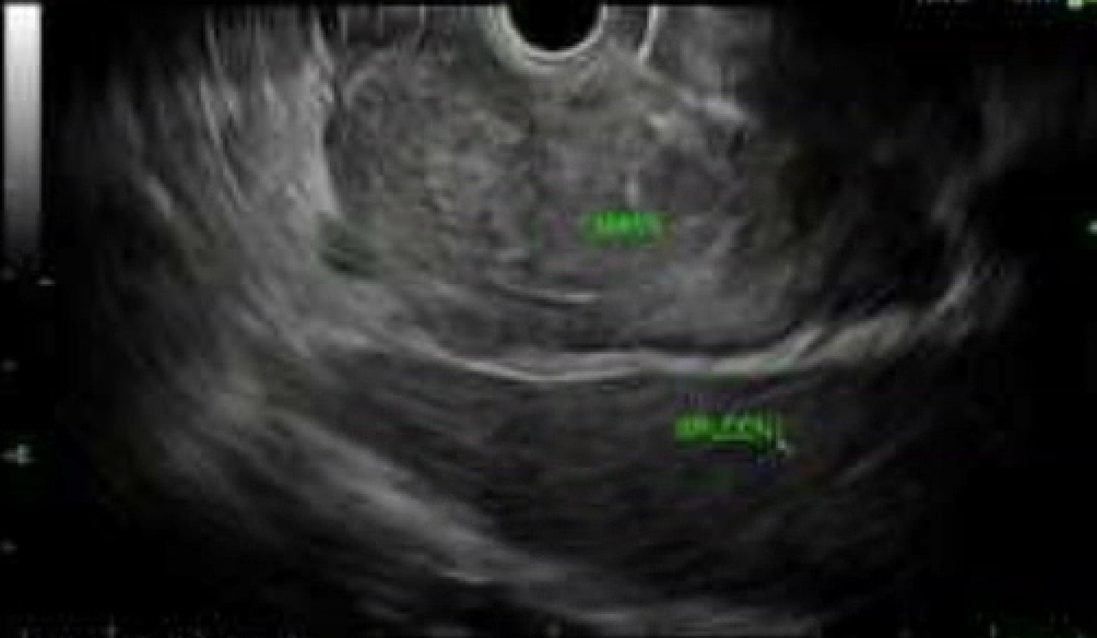
Figure: Endoscopic ultrasound (EUS) view of the gastric mass extending into the area of the spleen
Disclosures: Ross Dies indicated no relevant financial relationships. Farhan Mohiuddin indicated no relevant financial relationships. Brooke McVaney indicated no relevant financial relationships. Jessica Bordes indicated no relevant financial relationships.
Ross M. Dies, MD, MS1, Farhan Mohiuddin, MD2, Brooke McVaney, DO3, Jessica Bordes, MD1. P4218 - Something Sinister: A GIST Masquerading as a Gastric Abscess, ACG 2025 Annual Scientific Meeting Abstracts. Phoenix, AZ: American College of Gastroenterology.