St. Luke's University Health Network Bethlehem, PA
Frank Lin, DO, Ayah Obeid, MD, Ummul Zakia, MD, Kunal Bhagatwala, MD, Manoj Mittal, MD St. Luke's University Health Network, Easton, PA Introduction: Burkitt lymphoma (BL) is an aggressive non-Hodgkin lymphoma driven by MYC gene translocation. It has three subtypes: endemic, sporadic, and immunodeficiency associated. While BL often involves the ileocecal region, primary gastric Burkitt lymphoma (PGBL) is rare and accounts for a small subset of gastrointestinal lymphomas. Here, we present a 64-year-old male who presented with epigastric pain, melenic stools, blood loss anemia and found to have PGBL as a bleeding gastric ulcer. Given its aggressive course, early recognition and prompt chemotherapy are crucial for remission.
Case Description/
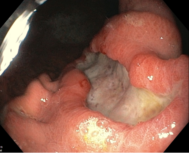
Methods: A 64-year-old male with past medical history significant for peptic ulcer disease initially presented to the emergency department with complaints of melena and epigastric pain for a duration of two to three days. CT abdomen showed findings suggestive of gastritis and a possible ulcer along the lesser curvature of the stomach with nonspecific low-grade lymphadenopathy involving the gastrohepatic lymph nodes. Esophagogastroduodenoscopy (EGD) showed a single large, irregular, and cratered malignant-appearing ulcer within the distal lesser body of the stomach measuring 3 x 10 cm (Figure 1). The pathology of the biopsied specimen showed a monotonous proliferation of neoplastic cells obliterating the normal gastric architecture with tingible body macrophages creating the classic “starry sky” appearance (Figure 2). Biopsy samples from the ulcer showed atypical proliferation of large B-cells with aberrant CD10 and BCL-6 expression, along with fluorescent in situ hybridization testing showing rearrangement of MYC;IgH t (8;14) (Figure 3).The patient was subsequently started on R-EPOCH chemotherapy. Discussion: Burkitt’s lymphoma with primary gastric manifestation can be a cause for ulcerative lesions and upper gastrointestinal bleeding. The two main histological subtypes of primary gastric Burkitt’s lymphoma include diffuse large B-cell lymphomas and MALT lymphomas. PGBL presents nonspecifically, often mimicking peptic ulcer disease, gastritis, or gastric adenocarcinoma. Symptoms include abdominal pain, nausea, gastrointestinal bleeding, or, in advanced cases, perforation. Diagnosis requires histopathology and immunohistochemistry due to variable endoscopic findings. One defining characteristic of Burkitt’s lymphoma is the chromosomal translocation between the Myc proto-oncogene and one of three immunoglobulin genes. Immediate intervention with R-EPOCH chemotherapy is recommended for five cycles.
Figure: Figure 1. A single large, irregular, and cratered malignant-appearing ulcer within the distal lesser body of the stomach measuring 3 x 10 cm.
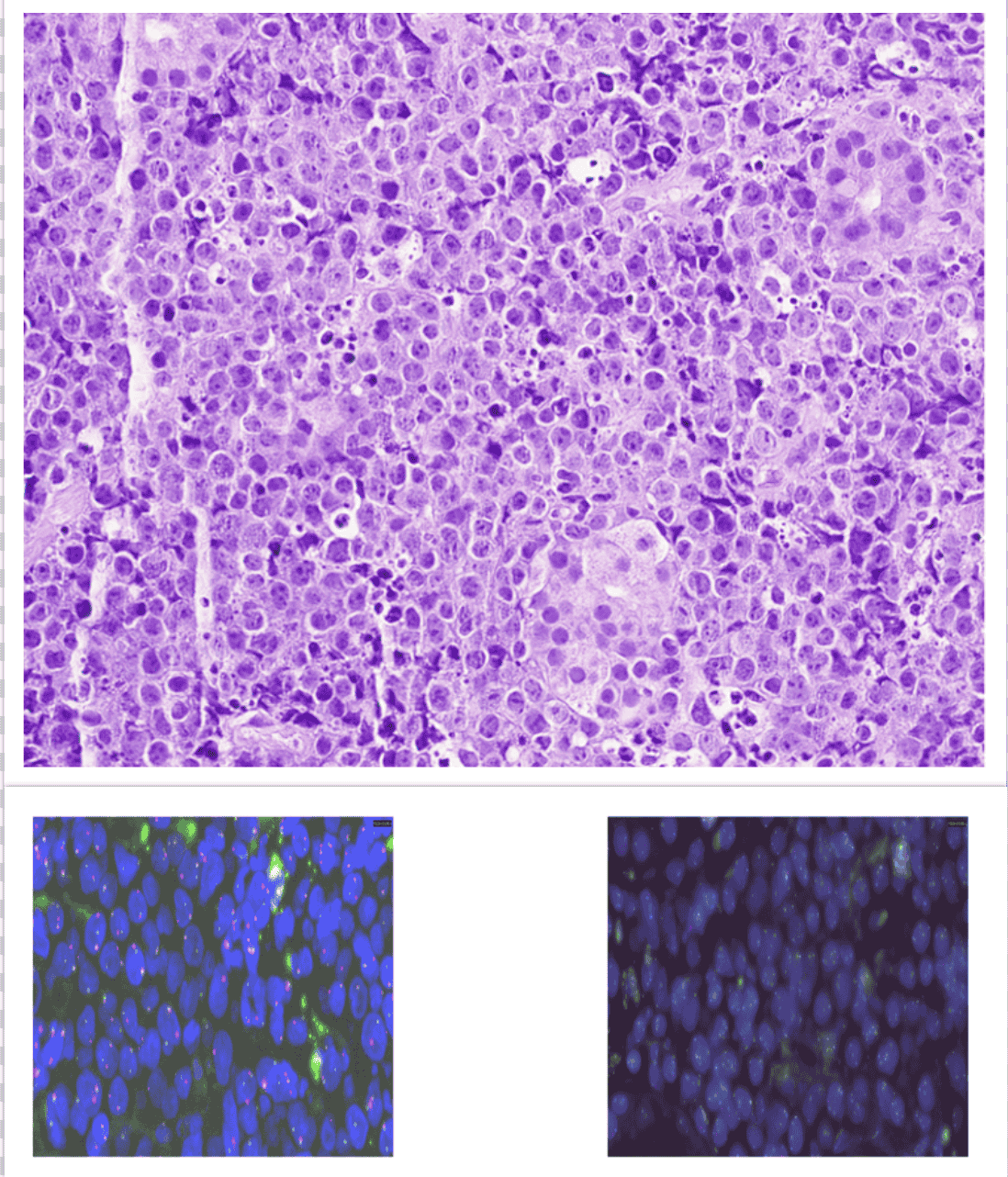
Figure: Figure 2. The neoplastic cells demonstrate obliteration of the normal gastric architecture. The cells are intermediate in size with round nuclei that are monotonous in appearance. Tingible body macrophages create the classic “starry sky” appearance.
Figure 3. Fluorescent in situ hybridization of the sample shows a MYC/IgH arrangement t(8;14), which is essential to the diagnosis of Burkitt’s lymphoma.
Disclosures: Frank Lin indicated no relevant financial relationships. Ayah Obeid indicated no relevant financial relationships. Ummul Zakia indicated no relevant financial relationships. Kunal Bhagatwala indicated no relevant financial relationships. Manoj Mittal indicated no relevant financial relationships.
Frank Lin, DO, Ayah Obeid, MD, Ummul Zakia, MD, Kunal Bhagatwala, MD, Manoj Mittal, MD. P4199 - An Unusual Cause of Melena: A Rare Case of Primary Gastric Burkitt’s Lymphoma Presenting as a Bleeding Gastric Ulceration, ACG 2025 Annual Scientific Meeting Abstracts. Phoenix, AZ: American College of Gastroenterology.