Silliman University Medical Center Foundation, Inc. Dumaguete, Negros Oriental, Philippines
Joyce Ramirez, MD1, Maria Joscel Torres, MD2, Paolo Macasaet, MD1, Dehuel Cuyacot, MD1 1Silliman University Medical Center Foundation, Inc., Dumaguete, Negros Oriental, Philippines; 2Negros Oriental Provincial Hospital, Dumaguete City, Negros Oriental, Philippines Introduction: Primary small bowel neoplasms are rare, accounting for 0.6% of total cancer cases and 3.3% of gastrointestinal malignancies. The majority of cases are adenocarcinoma (SBA) and are mostly found in the duodenum, making distal ileal involvement particularly rare, with an incidence of 7-17%. Diagnosis is often delayed due to vague symptoms and the lack of established screening protocols. The definitive diagnosis involves tissue sampling for histological and immunohistochemical analysis. To date, the incidence of small bowel cancers in the Philippines is only 0.3% per 100,000 population.
Case Description/
Methods: We report a rare case of a 76-year-old man diagnosed with primary small bowel adenocarcinoma (SBA) of the distal ileum, who initially presented with clinical features suggestive of acute appendicitis. When computed tomography (CT) imaging was done, it revealed an irregular mass in the right lower quadrant with infiltration of the distal ileum. Carcinoembryonic antigen (CEA) levels were within normal limits. The patient underwent segmental resection of the affected bowel. Histopathological analysis confirmed a non-ampullary adenocarcinoma with full-thickness invasion of the ileal wall. Immunohistochemical staining was positive for both cytokeratin 7 (CK7) and cytokeratin 20 (CK20), which favors a primary small bowel origin and rules out metastatic colorectal cancer. Based on these findings, the patient was diagnosed with stage IIA primary small bowel adenocarcinoma and was subsequently referred for adjuvant chemotherapy. Discussion: Small bowel adenocarcinoma (SBA) is rare, comprising less than 5% of GI malignancies. Its low incidence is linked to rapid transit, low microbial load, and mucosal immunity. Nonspecific symptoms often lead to delays in diagnosis, with most cases identified at advanced stages. CT imaging offers ~80% sensitivity for SBA, but tumors in the mid to distal ileum may be missed by standard endoscopy. Histopathology is essential for diagnosis, confirming glandular architecture and invasion depth. Immunohistochemical staining differentiates primary SBA from metastatic disease. Our patient’s tumor was staged IIA, invading the subserosa without nodal or distant spread. Surgical resection with lymphadenectomy remains the cornerstone of treatment. Though no standard chemotherapy exists, evidence supports adjuvant therapy in advanced stages. This case underscores the diagnostic complexity of SBA and the importance of imaging, histopathology, and timely intervention.
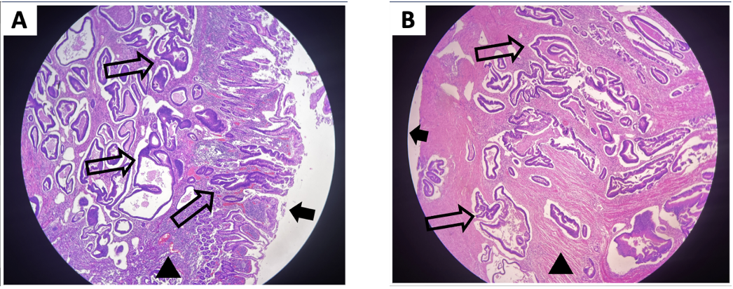
Figure: Periodic acid–Schiff–stained sample from the resected ileum as viewed in low-power magnification. (A) Neoplastic glands are seen at the mucosal surface and submucosa as indicated by the open arrows and arrowhead consecutively. (B) Neoplastic glands are seen infiltrating the muscularis propria as indicated by the arrowhead and extending to the serosa, indicated by the solid arrow.
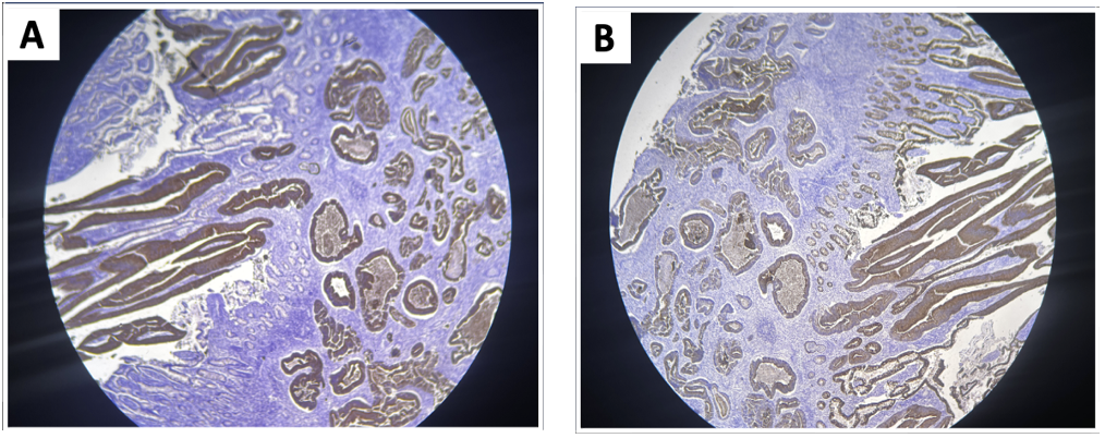
Figure: Immunohistochemical Staining of the resected ileum sample using (A) CK7 and (B) CK20, both showing a positive result.
Disclosures: Joyce Ramirez indicated no relevant financial relationships. Maria Joscel Torres indicated no relevant financial relationships. Paolo Macasaet indicated no relevant financial relationships. Dehuel Cuyacot indicated no relevant financial relationships.
Joyce Ramirez, MD1, Maria Joscel Torres, MD2, Paolo Macasaet, MD1, Dehuel Cuyacot, MD1. P4134 - Primary Adenocarcinoma of the Distal Ileum Presenting as Acute Appendicitis: A Rare Case From the Lower Gastrointestinal Tract, ACG 2025 Annual Scientific Meeting Abstracts. Phoenix, AZ: American College of Gastroenterology.

.jpg "Joyce Ramirez, MD photo")