Taofeek Kolade, MD, MBA, MPH1, Harleen Kaur, MD1, Meron Debesai, MD2 1Bayonne Medical Center, Bayonne, NJ; 2Carepoint Health/ Christ Hospital, Jersey City, NJ Introduction: Duodenal pseudomelanosis is a rare, benign condition characterized by dark pigmentation of the duodenal mucosa, typically discovered incidentally during esophagogastroduodenoscopy (EGD). Fewer than 150 cases have been reported. Histologically, it is defined by pigment-laden macrophages in the lamina propria, often containing iron sulfide. Although its exact pathogenesis remains unclear, it has been associated with chronic kidney disease, hypertension, diabetes mellitus, and the use of iron supplements or sulfur-containing medications. We present a case of duodenal pseudomelanosis in an elderly patient with concurrent Helicobacter pylori infection, a co-occurrence not previously reported and of uncertain significance.
Case Description/
Methods: A 76-year-old female with hypothyroidism, hypertension, and hyperlipidemia presented with intermittent reflux, bloating, and gas. She denied NSAID or iron supplement use. EGD revealed mild gastritis, two superficial antral ulcers, five gastric polyps, and patchy dark pigmentation in the second portion of the duodenum. Gastric biopsies were positive for H. Pylori in both the body and antrum. The polyps were resected and identified as tubular adenomas. Duodenal biopsies showed pigment-laden macrophages in the lamina propria consistent with pseudomelanosis, without iron staining, dysplasia, malignancy, or active inflammation. Discussion: This case highlights duodenal pseudomelanosis as a benign, incidental finding in an elderly patient undergoing evaluation for dyspepsia. While the condition is often associated with iron supplementation, its absence here suggests that chronic antihypertensive therapy and advanced age may have contributed to pigment deposition. The concurrent presence of Helicobacter pylori infection adds interest, as no prior association with pseudomelanosis has been reported. Although a causal relationship cannot be established from a single case, this co-occurrence raises the possibility of a novel interaction warranting further observation. Recognizing pseudomelanosis as a non-threatening entity is crucial to avoid unnecessary interventions. This case underscores the importance of interpreting endoscopic findings within clinical and histologic context to guide appropriate management.
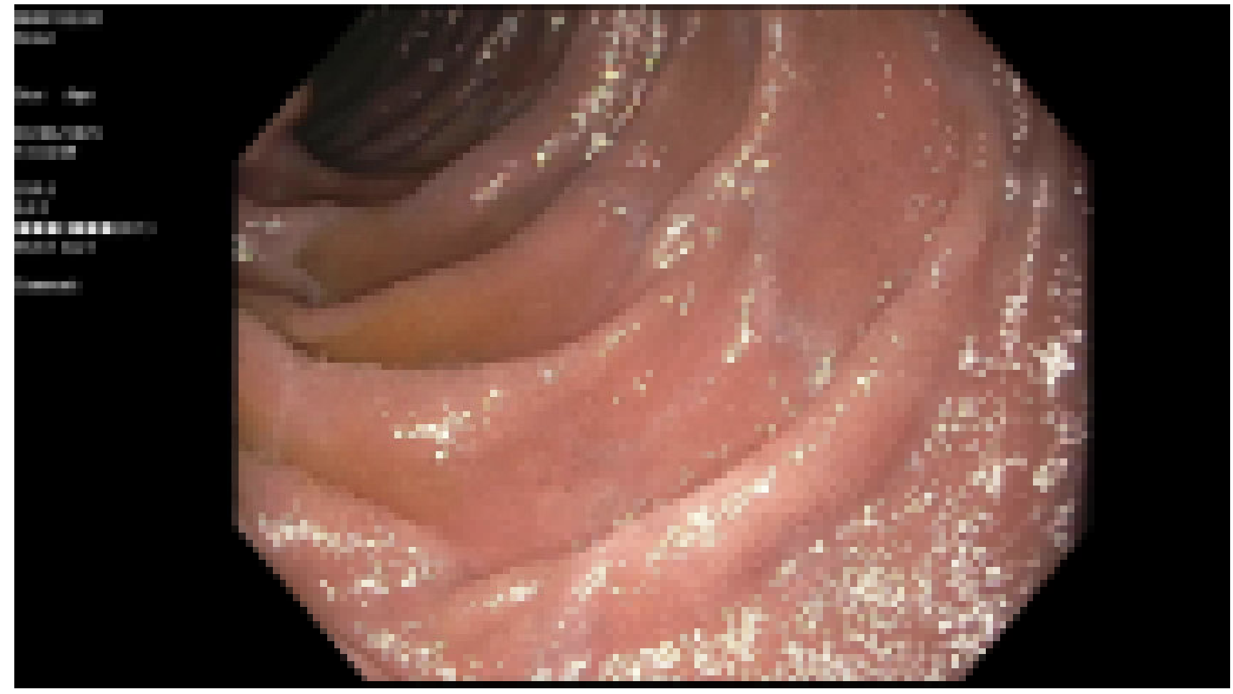
Figure: Figure 1. Patchy mild mucosal changes characterized by discoloration were found in the duodenal bulb
Disclosures: Taofeek Kolade indicated no relevant financial relationships. Harleen Kaur indicated no relevant financial relationships. Meron Debesai indicated no relevant financial relationships.
Taofeek Kolade, MD, MBA, MPH1, Harleen Kaur, MD1, Meron Debesai, MD2. P4132 - An Incidental Finding of Duodenal Pseudomelanosis With Concurrent <i>H. pylori</i>, ACG 2025 Annual Scientific Meeting Abstracts. Phoenix, AZ: American College of Gastroenterology.