Staten Island University Hospital, Northwell Health Staten Island, NY
Chloe Lahoud, MD, Mark Tawfik, DO, Ahmed Elfiky, MD, Harika Kandlakunta, MD, Jean Chalhoub, MD Staten Island University Hospital, Northwell Health, Staten Island, NY Introduction: Gangliocytic paraganglioma (GP) is a rare tumor that commonly involves the gastrointestinal system and often occurs in the second portion of the duodenum. Aside from gastrointestinal involvement, some case reports present GP of the lungs, heart, head and neck. GP is characterized by its triphasic cellular differentiation on histopathology. Most GPs are considered benign and are amenable to excision, however, periampullary GPs should be considered as a tumor with malignant potential.
Case Description/
Methods: This is the case of a 73-year-old female presenting for an unintentional 10-pound weight loss over the previous year and a computed tomography (CT) scan of the abdomen and pelvis showing a mass in the second portion of the duodenum with omental nodules. She had no other symptoms. An upper endoscopy revealed a pedunculated 5 cm polypoid mass that was endoscopically resected and found to be a gangliocytic paraganglioma on pathology. GP is the third most frequent histopathologic type of gastrointestinal neuroendocrine tumors (NET) after gastrinomas and somatostatinomas. Clinical presentations vary from asymptomatic patients to others having gastrointestinal bleeding, melena, anemia and abdominal pain. GP presents as a single solid well-demarcated mass that is polypoid, pedunculated or sessile. Histological features of GP show three cell types: spindle cells, ganglion or ganglion-like cells, and epithelioid cells. Biopsy specimens might not contain all 3 characteristic cell types, thus definitive diagnosis could be challenging. Periampullary GP should be considered as a tumor with malignant potential. The optimal treatment has not been clarified, but endoscopic resection remains the most common treatment of choice. Discussion: This case underscores the importance of including rare tumors with malignant potential, such as gangliocytic paraganglioma (GP), in the differential diagnosis of duodenal masses. Histologic features, including spindle, ganglion, or epithelioid cells, combined with characteristic immunohistochemical staining, are essential for confirmation. Treatment typically involves endoscopic or surgical resection, depending on the depth of invasion. Further longitudinal studies are needed to establish standardized management protocols and enhance patient care.
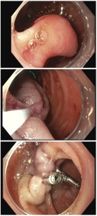
Figure: Fig 1: A pedunculated 5cm polypoid mass in the second portion of the duodenum (gangliocytic paraganglioma) (Fig 1A) removed endoscopically with a hot snare (Fig 1B) followed by the placement of 3 endoclips (Fib 1C).
Disclosures: Chloe Lahoud indicated no relevant financial relationships. Mark Tawfik indicated no relevant financial relationships. Ahmed Elfiky indicated no relevant financial relationships. Harika Kandlakunta indicated no relevant financial relationships. Jean Chalhoub indicated no relevant financial relationships.
Chloe Lahoud, MD, Mark Tawfik, DO, Ahmed Elfiky, MD, Harika Kandlakunta, MD, Jean Chalhoub, MD. P4123 - Case of a Gangliocytic Paraganglioma of the Duodenum Presenting as Isolated Weight Loss: Diagnostic Challenges and Management Highlights, ACG 2025 Annual Scientific Meeting Abstracts. Phoenix, AZ: American College of Gastroenterology.