Aaron Goffinet, MD1, Meredith Yellen, MD2, Edwin McDonald, MD1 1University of Chicago Medical Center, Chicago, IL; 2University of Chicago, Chicago, IL Introduction: Monomorphic epitheliotropic intestinal T-cell lymphoma (MEITL) is a rare and aggressive primary intestinal lymphoma, formerly classified as enteropathy associated T-cell lymphoma (EATL) type II. Reclassified by the WHO in 2017, MEITL lacks the celiac disease association seen in EATL type I and is far less common. Due to the aggressive nature of this disease, diagnosis is most often made via surgical intervention in an emergent fashion due to intestinal perforation or obstruction. In one of the largest cohort studies of MEITL to date, 30 of 35 patients were diagnosed surgically and only 5 identified endoscopically. We present a case of a patient with subacute onset abdominal symptoms diagnosed with MEITL via double-balloon enteroscopy (DBE).
Case Description/
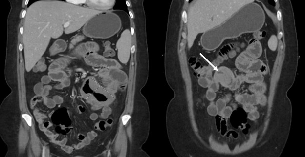
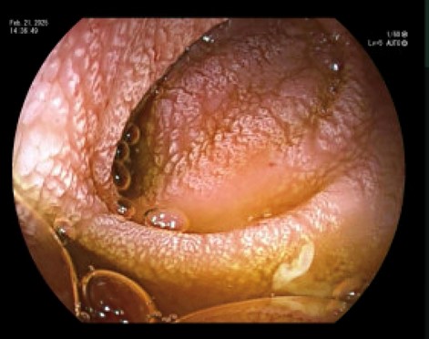
Methods: A 54-year-old Hispanic woman with a history of iron deficiency anemia (IDA) refractory to IV iron supplementation presented with subacute onset of progressive abdominal pain, early satiety, and 20-pound weight loss. She had an EGD and colonoscopy three years prior to presentation for the IDA which were both overall normal. Celiac serologies were also negative. Upon presentation CT scan (Figure 1) demonstrated thickened jejunal loops and borderline mesenteric lymphadenopathy. Given jejunal pathology on imaging, decision made to pursue DBE which reveal severe mucosal changes (Figure 2) in distal duodenum and jejunum. Histology from biopsies demonstrated dense monomorphic lymphoid cell infiltration and CD3, CD2, CD7, CD8, and CD56 positivity on IHC consistent with MEITL. Discussion: MEITL is a rare but severe form of intestinal lymphoma most diagnosed in Asian and Hispanic patients. There is limited literature which describes endoscopic diagnosis of MEITL, and to a lesser extent the use of DBE for diagnosis. The median onset of survival is just 7 months; however, early diagnosis can lead to significant improvement in mortality. Our case is a rare presentation which demonstrated early endoscopic evaluation using DBE with prompt diagnosis and initiation of therapy. Our patient has remained without need for operative intervention and on PET imaging has demonstrated partial response to her chemotherapy regimen.
References: Min GJ, Oh YE, Jeon Y, et al. Hematopoietic stem cell transplantation to improve prognosis in aggressive monomorphic epitheliotropic intestinal T-cell lymphoma. Front Oncol. 2024;14:1388623. Published 2024 Nov 21.
Figure: Figure 1: Multi-focal short segment jejunal bowel wall thickening seen on initial CT upon presentation. Greatest diameter of dilation was 5.1 cm of an 8 cm segment.
Figure: Figure 2: Segmental severe mucosal changes characterized by atrophy, granularity, nodularity, and scalloping. This pattern was seen largely in the fourth portion of the duodenum, jejunum, and proximal ileum.
Disclosures: Aaron Goffinet indicated no relevant financial relationships. Meredith Yellen indicated no relevant financial relationships. Edwin McDonald indicated no relevant financial relationships.
Aaron Goffinet, MD1, Meredith Yellen, MD2, Edwin McDonald, MD1. P4120 - Monomorphic Epitheliotropic Intestinal T-Cell Lymphoma Diagnosed with Double-Balloon Enteroscopy, ACG 2025 Annual Scientific Meeting Abstracts. Phoenix, AZ: American College of Gastroenterology.