Arti Patel, MD1, Rohit Khanna, DO2, Ahmed Deabes, MD3 1Eisenhower Medical Center, Rancho Mirage, CA; 2Scripps Green Hospital, La Jolla, CA; 3Scripps Clinic/Scripps Green Hospital, La Jolla, CA Introduction: Multicystic peritoneal mesothelioma (MCPM), a rare variant of peritoneal mesothelioma, is characterized by fluid-filled cysts lined by mesothelial cells. As of 2017, just over 200 cases have been reported worldwide. Its annual incidence is 2 cases per million, mostly affecting reproductive-age women. Risks are chronic inflammation and prior abdominal surgeries, though studies suggest neoplastic potential due to rare risk of malignant transformation. Vague and insidious symptoms can delay diagnosis. We report a case of malignant peritoneal mesothelioma (MPM) in a 65-year-old male, with features suggesting malignant transformation of MCPM.
Case Description/
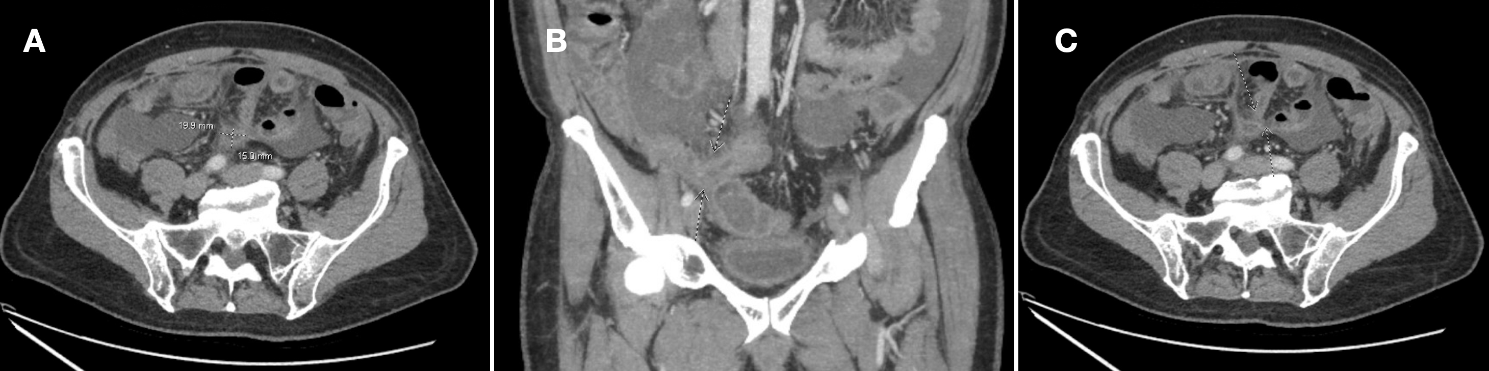
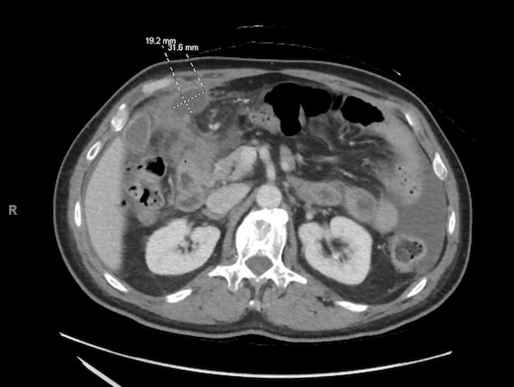
Methods: A 65-year-old male with benign prostatic hyperplasia, laparoscopic appendectomy and chronic prostatitis presented with severe acute-on-chronic right-sided and periumbilical pain, along with one month of anorexia, fatigue, and unintentional 10-pound weight loss. His vital signs were stable. Labs were WBC 15.3 K/mcL, INR 1.5, CRP 261.7 mg/L, ESR 59 mm/hr and ferritin 2211 ng/mL. A CT abdomen and pelvis with Enterography showed moderate ascites and loculated rim-enhancing peritoneal fluid collections (Figure 1) into the gallbladder fossa and small bowel mesentery, concerning for an entero-enteric fistula (Figure 2). Additionally, sigmoid colon and terminal ileum thickening raised concerns for inflammatory bowel disease. Comprehensive infectious, autoimmune, and malignancy workup, endoscopy and colonoscopy were negative. Surgery consultation recommended open laparotomy given persistent symptoms which revealed diffuse abdominal ascites, pelvic and hepatic flexure fluid collections, omental caking, peritoneal implants and friable adhesions, with intraoperative omental biopsy confirming biphasic MPM. Discussion: Unlike pleural mesothelioma, MCPM is infrequently linked to asbestos exposure. Though the etiology is unclear and primarily idiopathic, radiation, chronic inflammation, and familial cancer syndromes likely play pathogenic roles. Beyond its etiological ambiguity, diagnosis is challenging as symptoms of MCPM mimic other intra-abdominal processes. Imaging typically shows solid features, such as mass-like lesions, nodules and omental caking, none of which were identified in this patient's CT scan. This case highlights the rare malignant transformation of MCPM and its diagnostic difficulties. Further research is crucial to elucidate the malignant potential of MCPM and to establish comprehensive diagnostic and management guidelines.
Figure: Figure 1. A discrete, thin rim-enhancing fluid collection along the anterior/inferior margin of the left liver lobe measuring 1.9 x 3.2 x 3.9 cm.
Figure: Figure 2. A. An inferior small bowel mesenteric fluid collection, 1.5 x 2.0 cm in transverse dimension, with slight peripheral enhancement with B. communication with the distal ileum and C. with mid abdominal small bowel loops concerning for an entero-enteric fistula.
Disclosures: Arti Patel indicated no relevant financial relationships. Rohit Khanna indicated no relevant financial relationships. Ahmed Deabes indicated no relevant financial relationships.
Arti Patel, MD1, Rohit Khanna, DO2, Ahmed Deabes, MD3. P4119 - Atypical Presentation of Malignant Peritoneal Mesothelioma With Cystic Features in a Male Patient, ACG 2025 Annual Scientific Meeting Abstracts. Phoenix, AZ: American College of Gastroenterology.