Nisar Amin, MD1, M Housam Nanah, MD2, Alaa Habash, MD1, Harleen Chela, MD1, Ebubekir Daglilar, MD1 1Charleston Area Medical Center, Charleston, WV; 2Cleveland Clinic, Cleveland, OH Introduction: Hepatoduodenal fistulas are rare connections between the liver and duodenum, typically arising from liver abscesses, tumors, gastric perforation, or iatrogenic causes such as transarterial chemoembolization. We present a case of a hepatoduodenal fistula secondary to a peptic ulcer, presenting with severe gastrointestinal bleeding and a complicated clinical course.
Case Description/
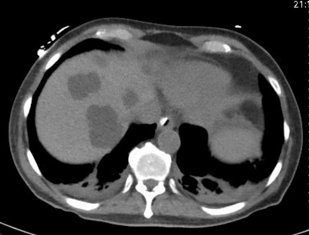
Methods: A 76-year-old male with a history of atrial fibrillation and CVA on Plavix, and multiple comorbidities was transferred from an outside facility after presenting with general malaise. He had been previously diagnosed with a presumed contained perforated duodenal ulcer (without endoscopy) and discharged on oral PPI therapy. He subsequently returned with large-volume hematochezia, syncope, and hemoglobin of 5.4 g/dL, along with an elevated BUN-to-creatinine ratio and epigastric pain. He had a recent history of excessive NSAID use. On physical exam he had mild diffuse abdominal tenderness and melena on rectal examination. Initial abdominal CT revealed a suspected contained duodenal perforation, necrotic hepatic mass, and possible duodeno-hepatic fistula at the porta hepatis(Fig.1).EGD showed a large duodenal clot, a perforated ulcer with a fistula into hepatic parenchyma, multiple adherent clots, and two non-bleeding angiectasias(Fig.2). Given the ulcer size and fistula, surgery was advised. Exploratory laparotomy confirmed duodenal perforation into a bleeding hepatic cyst. Surgical procedures included gastroplasty, pyloric exclusion, Roux-en-Y gastrojejunostomy, marsupialization of the cyst, fistula takedown, Graham patch, omental pedicle flap, and open cholecystectomy. Postoperative CT showed a worsening gallbladder fossa collection and high bilious drain output, indicating a duodenal stump leak. After failed transhepatic cholangiography, IR performed biliary diversion. The patient improved on antibiotics and was discharged. Discussion: Gastrointestinal perforations are a significant cause of morbidity and mortality, usually presenting with bleeding. Fistulization to adjacent organs may contain bleeding and delay diagnosis. Hepatoduodenal fistulas often cause nonspecific symptoms—abdominal pain, fever, jaundice—or are found incidentally. Complications include biliary sepsis, infection, or GI bleeding. Diagnosis typically involves imaging (CT, MRCP, cholangiography) and sometimes endoscopy. Management varies by case, depending on stability and complications, and may be conservative, endoscopic, or surgical.
Figure: Duodenal perforation, necrotic hepatic mass, and possible duodeno-hepatic fistula at the porta hepatis.
Figure: Large duodenal clot, a perforated ulcer with a fistula into hepatic parenchyma, multiple adherent clots, and non-bleeding angiectasias.
Disclosures: Nisar Amin indicated no relevant financial relationships. M Housam Nanah indicated no relevant financial relationships. Alaa Habash indicated no relevant financial relationships. Harleen Chela indicated no relevant financial relationships. Ebubekir Daglilar indicated no relevant financial relationships.
Nisar Amin, MD1, M Housam Nanah, MD2, Alaa Habash, MD1, Harleen Chela, MD1, Ebubekir Daglilar, MD1. P4115 - A Rare Case of Hepatoduodenal Fistula Secondary to Duodenal Ulcer Disease, ACG 2025 Annual Scientific Meeting Abstracts. Phoenix, AZ: American College of Gastroenterology.