Raya Alashram, MD, Ahmad Abulawi, MD, Stephen Hasak, MD, MPH, Seth Richter, MD, FACG Albany Medical Center, Albany, NY Introduction: Cytomegalovirus (CMV) is a common opportunistic infection, particularly affecting immunocompromised individuals. CMV can involve any segment of the gastrointestinal (GI) tract, with the esophagus and colon being most frequently affected. Small bowel involvement, especially of the jejunum, is rare and often underrecognized. When present, CMV enteritis can result in significant morbidity and necessitates early diagnosis and prolonged antiviral therapy to prevent serious complications. We present a case of jejunal CMV ulceration in a kidney transplant recipient on chronic immunosuppression.
Case Description/
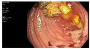
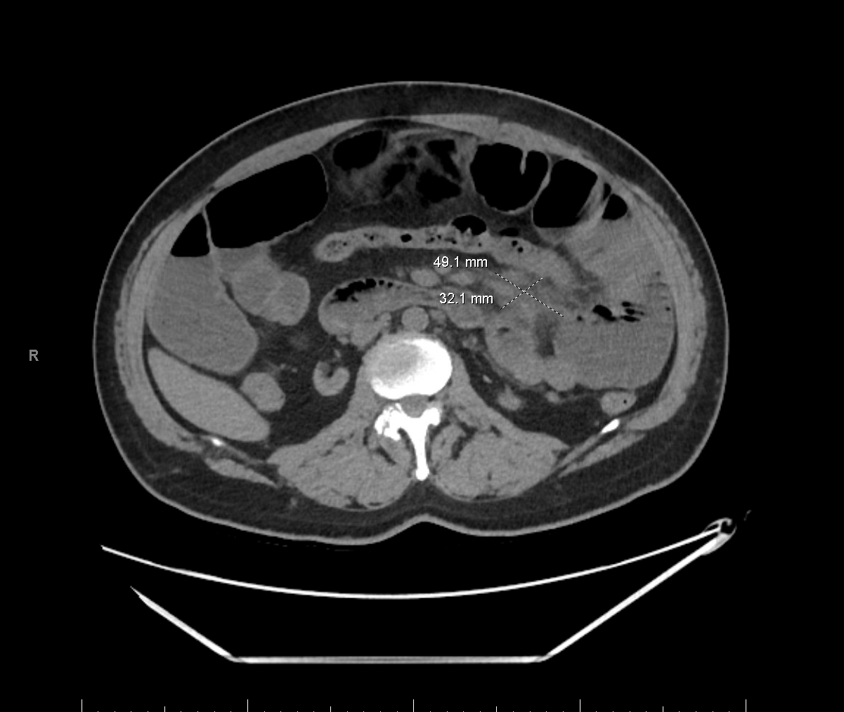
Methods: A 46-year-old male with focal segmental glomerulosclerosis, status post-deceased donor kidney transplant (DDKT), maintained on tacrolimus and belatacept, presented with worsening sharp upper abdominal pain, bloating, nausea, diarrhea, anorexia, and a 35-pound weight loss over three months. Initial computed tomography imaging revealed enteritis and features suggestive of small bowel obstruction (Figure 1). Esophagogastroduodenoscopy (EGD) was unremarkable, and biopsies from the stomach and duodenum were inconclusive. Due to persistent symptoms, push enteroscopy was performed, which revealed ulceration in the jejunum (Figure 2). Biopsy of the lesion confirmed active CMV enteritis. The terminal ileum and colon appeared normal. The patient was initiated on intravenous ganciclovir by the infectious disease team, leading to symptomatic improvement. Discussion: Jejunal CMV ulceration is a rare manifestation of tissue-invasive CMV disease, typically seen in immunocompromised hosts such as transplant recipients. Intravenous ganciclovir is the preferred initial treatment, often followed by oral valganciclovir for maintenance. Management may require adjustment of immunosuppressive therapy to enhance viral clearance. Importantly, serum CMV PCR levels may not correlate with mucosal disease activity, underscoring the need for tissue diagnosis. Delay in recognition can result in severe complications, including stricture, bleeding, or perforation. This case highlights the importance of considering CMV enteritis in transplant recipients with persistent GI symptoms and supports the utility of push enteroscopy for diagnosis when conventional EGD is inconclusive.
Figure: Figure 1 CT abdomen showing enteritis and features suggestive of small bowel obstruction
Figure: Figure 2 Clean base ulcer in the jejunum
Disclosures: Raya Alashram indicated no relevant financial relationships. Ahmad Abulawi indicated no relevant financial relationships. Stephen Hasak: Castle Biosciences – Speakers Bureau. Conmed – Consultant. Seth Richter indicated no relevant financial relationships.
Raya Alashram, MD, Ahmad Abulawi, MD, Stephen Hasak, MD, MPH, Seth Richter, MD, FACG. P4084 - Beyond the Colon: Rare Jejunal CMV Ulcer in an Immunosuppressed Patient, ACG 2025 Annual Scientific Meeting Abstracts. Phoenix, AZ: American College of Gastroenterology.