Dylan Vainer, MD1, Mehul Trivedi, MD2, Nicole Leonard, MD2, Valarie McMurtry, MD, PhD2, Juan F. Gallegos-Orozco, MD, FACG2 1University of Utah, Salt Lake City, UT; 2University of Utah Health, Salt Lake City, UT Introduction: Choroidal melanoma is a rare type of melanoma that occurs within the uveal tract of the eye, with an incidence rate of about 1,500 cases per year in the United States. The liver is the most common site of metastases, however acute liver failure secondary to hepatic infiltration is extremely rare and there are only a few reported cases in the literature. We report a unique case of a woman who presented with acute liver failure secondary to metastatic choroidal melanoma despite successful ocular radiation treatment two years prior.
Case Description/
Methods: The patient was a 43-year-old female with a history of choroidal melanoma that was treated with plaque brachytherapy two years prior. She had been scheduled to undergo biannual surveillance abdominal imaging however was lost to follow-up. She presented with several weeks of worsening abdominal pain, distention, constipation, and anorexia. Her initial labs were notable for AST 199, ALT 62, total bilirubin 1.6, and ALP 44. CT imaging demonstrated hepatomegaly with right portal vein and hepatic vein thrombus, moderate ascites, and a 1.4 cm hypoenhancing lesion adjacent to the caudate lobe. Analysis of ascitic fluid revealed a SAAG of 2.1, consistent with portal hypertension. Viral, autoimmune, and genetic causes of liver injury were ruled out. Over the next two weeks, her clinical condition deteriorated and her labs worsened: AST 320, ALT 86, total bilirubin 9.9, Na 127, albumin 2.4, and INR 2.6. A repeat MRI abdomen revealed new sub-centimeter lesions throughout the liver and a few peripheral wedge-shaped lesions that were favored to represent infarcts. A trans-jugular liver biopsy showed loosely cohesive cells with high nuclear to cytoplasmic ratios, consistent with metastatic melanoma. The patient became increasingly encephalopathic and lethargic over the next several days. Unfortunately, the patient’s metastatic cancer and poor clinical condition precluded liver transplant candidacy or systemic therapy. Aggressive measures were withdrawn and she expired shortly thereafter. Discussion: Acute liver failure secondary to diffuse involvement of choroidal melanoma is very rare and has been described only a handful of times. This patient’s worsening liver function was secondary to both diffuse hepatic melanoma infiltration and ischemia related to tumor-related necrosis or portal and hepatic vein thrombus. This case highlights that metastases should be considered in a patient with a history of choroidal melanoma and new-onset liver decompensation.
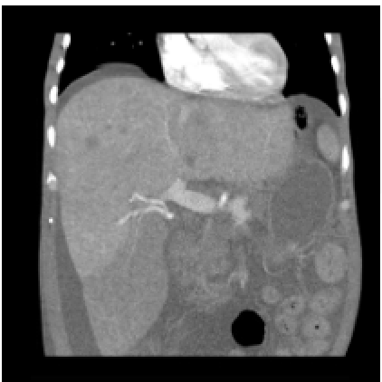
Figure: Hepatomegaly with marked heterogeneity of the liver and multiple indeterminate lesions and right portal vein thrombus
Figure: Liver biopsy preparation shows loosely cohesive malignant cells with high nuclear to cytoplasmic ratios, prominent nucleoli, and associated pigment. Core biopsies demonstrate nested sheets of these cells within scattered entrapped benign hepatocytes.
Disclosures: Dylan Vainer indicated no relevant financial relationships. Mehul Trivedi indicated no relevant financial relationships. Nicole Leonard indicated no relevant financial relationships. Valarie McMurtry indicated no relevant financial relationships. Juan Gallegos-Orozco: Gilead – Grant/Research Support. Hanmi – Grant/Research Support. Intercept – Grant/Research Support. Mirum – Grant/Research Support.
Dylan Vainer, MD1, Mehul Trivedi, MD2, Nicole Leonard, MD2, Valarie McMurtry, MD, PhD2, Juan F. Gallegos-Orozco, MD, FACG2. P4007 - Acute Liver Failure Secondary to Metastatic Choroidal Melanoma, ACG 2025 Annual Scientific Meeting Abstracts. Phoenix, AZ: American College of Gastroenterology.