Jose Garcia-Corella, MD1, Shyam Patel, MD2, Carol Singh, MBBS3, Sindhu Chadalawada, MD4, Marina Roytman, MD5 1UCLA-Kern Medical Center, Bakersfield, CA; 2California Pacific Medical Center, San Francisco, CA; 3Dayanand Medical College and Hospital, New Jersey, NJ; 4UCSF Fresno, Fresno, CA; 5Hepatology Fellowship, UCSF Fresno, Fresno, CA Introduction: Primary squamous cell carcinoma of the liver (PSCCL) is an exceptionally rare malignancy, with only a few cases documented in literature. Its pathogenesis remains unclear, as the liver lacks native squamous epithelium. It is hypothesized to arise from malignant transformation of squamous metaplasia from chronic inflammation, pre-existing hepatic cysts or abscesses. Due to its rarity, PSCCL often poses diagnostic challenges and lacks standardized treatment guidelines. We report a case of PSCCL in a previously healthy male, highlighting the clinical presentation, diagnostic approach and therapeutic considerations.
Case Description/
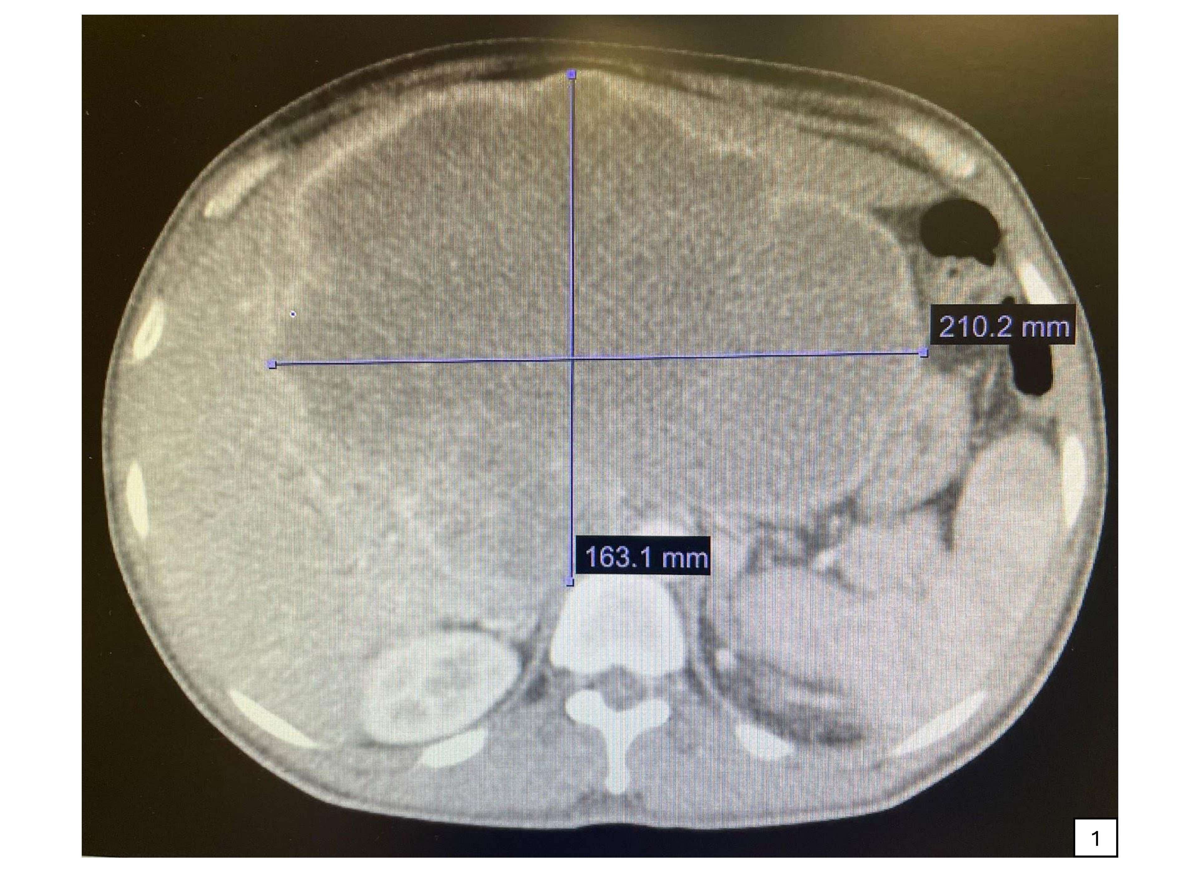
Methods: A 45-year-old otherwise healthy Hispanic man presented with right upper quadrant pain associated with night sweats and weight loss. Laboratories were notable for normocytic anemia, hypoalbuminemia, alkaline phosphatase 389 U/L, alanine aminotransferase 40 U/L, and aspartate aminotransferase 57 U/L. CT scan of the abdomen/pelvis demonstrated a 21x16 cm peripherally-enhancing liver mass with retroperitoneal lymphadenopathy. Tumor markers, including alpha fetoprotein, carcinoembryonic antigen, and carbohydrate antigen 19-9, were within normal limits. Endoscopy and colonoscopy were performed, no obvious mass was identified. Biopsy of the liver mass was consistent with moderately differentiated squamous cell carcinoma as evidenced by positive cytokeratin 5/6 and p40 immunohistochemical staining. Given the aggressive nature of the malignancy, he received a round of carboplatin/gemcitabine prior to discharge. He is to follow with oncology and hepatobiliary surgery. Discussion: PSCCL is a rare and aggressive malignancy, with no identifiable risk factors and a variety of nonspecific clinical and biochemical findings at presentation, as described by a limited number of case reports. The continuous peripheral enhancement seen on single-phase CT scan is frequently found on malignant lesions, including metastasis, but it is not specific for a single etiology. The diagnosis was ultimately made by pathologic analysis with immunohistochemistry, as has been the case with all previously reported PSCCL. Definitive treatment is surgical, with complete excision of the tumor being the goal, however, large masses or inoperable cases benefit from adjuvant chemotherapy or radiotherapy. Prognosis is poor, and reported to be worse in the presence of liver cysts. Though rare, this case highlights the importance of clinical suspicion and a thorough evaluation of patients with liver lesions.
Figure: Figure 1. 21 x 16 cm peripherally-enhancing liver mass demonstrated on computed tomography scan of the abdomen/pelvis.
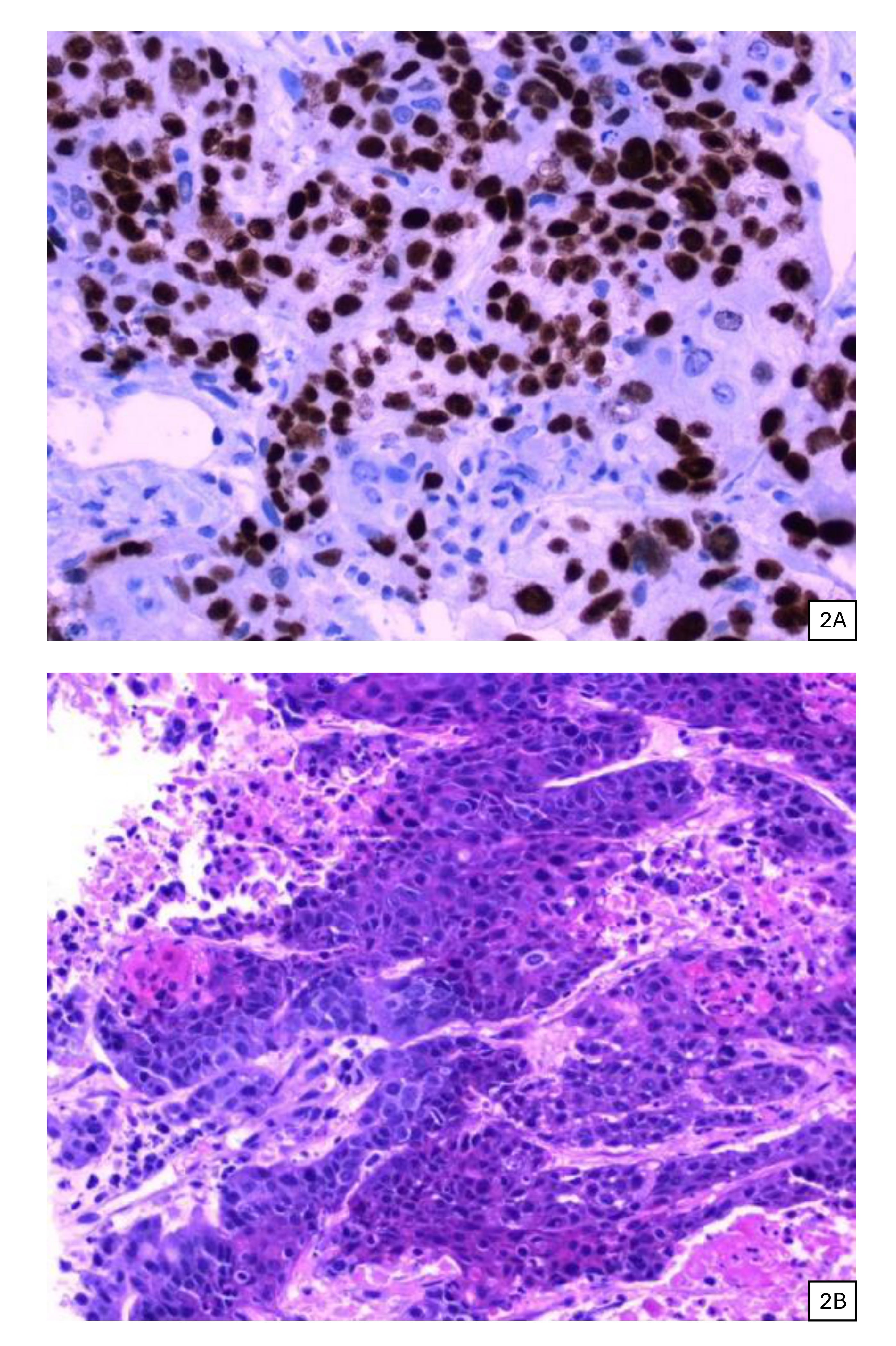
Figure: Figures 2A. Liver tissue with positive p40 immunohistochemistry supporting squamous differentiation; 2B. Liver squamous cell carcinoma with necrosis and keratin production.
Disclosures: Jose Garcia-Corella indicated no relevant financial relationships. Shyam Patel indicated no relevant financial relationships. Carol Singh indicated no relevant financial relationships. Sindhu Chadalawada indicated no relevant financial relationships. Marina Roytman: Gilead – Speakers Bureau. Ipsen – Advisory Committee/Board Member. Madrigal – Speakers Bureau. Salix – Speakers Bureau.
Jose Garcia-Corella, MD1, Shyam Patel, MD2, Carol Singh, MBBS3, Sindhu Chadalawada, MD4, Marina Roytman, MD5. P3930 - Hepatic Squamous Cell Carcinoma: A Rare Primary Malignancy, ACG 2025 Annual Scientific Meeting Abstracts. Phoenix, AZ: American College of Gastroenterology.

 photo")