The Warren Alpert Medical School of Brown University Warwick, RI
Gurjot Singh, DO1, Tamara Koritarov, DO, MSc2, Angela Fishman, DO3 1The Warren Alpert Medical School of Brown University, Warwick, RI; 2Kent Hospital, Providence, RI; 3Kent Hospital, Warwick, RI Introduction: The incidence rate of esophageal cancer and neuroendocrine tumors, as per the National Cancer Institute (NCI), are both reported to be approximately 1% of all cancer cases diagnosed. This case explores the unlikely diagnosis of an individual who was found to have both esophageal and neuroendocrine tumors as a primary malignancy during a hospitalization event.
Case Description/
Methods: A 51 year old male with a past medical history of alcohol use disorder, OSA, depression, GERD, and iron deficiency anemia presented for alcohol withdrawal. The patient endorsed right upper quadrant pain which had been intermittently occurring for the past four months, fatigue, and lightheadedness. On admission, the patient was sinus tachycardic with all other vitals stable. CT of the chest, abdomen, and pelvis demonstrated incidental marked circumferential thickening in the distal esophageal wall, enlarged left celiac lymph nodes concerning for metastatic adenopathy, and a suspected 4.3 x 3.4 cm right hepatic lobe lesion, concerning for metastatic disease. EGD was performed and demonstrated a partially obstructing, friable, granular, and nodular mass at the distal esophagus. Pathology was found to demonstrate poorly differentiated adenocarcinoma. Ultrasound of the right upper quadrant demonstrated numerous hepatic masses up to 5.8 cm in the right hepatic lobe. Liver biopsy was completed with pathology demonstrating well-differentiated neuroendocrine tumor. The patient was referred to oncology for PET scan completion and treatment. In addition, the patient was referred to thoracic surgery for evaluation and treatment of esophageal cancer. Discussion: Esophageal and neuroendocrine tumors are both rare processes with a low incidence in the general population. The co-existence of these two pathologic processes are rarely reported in literature. Although the cancers may be distinct histologically, this case entertains the possibility of ascertainment bias where neuroendocrine tumors may be more easily identified than esophageal cancer. In the case above, the esophageal wall thickening was an incidental finding when the main area of concern was the patient’s abdominal pain on presentation. Further research and reporting may be needed to provide a clearer relation between the two tumors.
Figure: Computed tomography (CT) of the abdomen and pelvis (A.) axial view of the 4.3 x 3.4 cm right hepatic lobe lesion (B.) coronal view of the right hepatic lobe lesion.
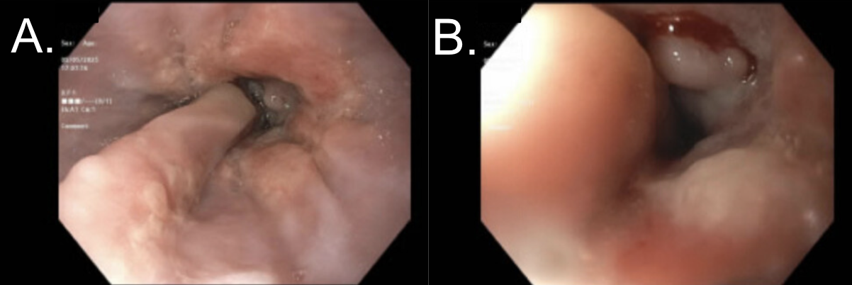
Figure: EGD findings of (A.) lower third of the esophagus with a mass at 35 cm (B.) lower third of the esophagus demonstrating a partially obstructing mass with the scope unable to traverse at 40 cm.
Disclosures: Gurjot Singh indicated no relevant financial relationships. Tamara Koritarov indicated no relevant financial relationships. Angela Fishman indicated no relevant financial relationships.
Gurjot Singh, DO1, Tamara Koritarov, DO, MSc2, Angela Fishman, DO3. P3925 - Lightning Strikes Twice: A Rare Case of Esophageal Adenocarcinoma and Neuroendocrine Tumor Co-Diagnosis, ACG 2025 Annual Scientific Meeting Abstracts. Phoenix, AZ: American College of Gastroenterology.